
If you’ve ever peered into a drop of water from a mossy roof or garden and spotted a slow-moving, eight-legged creature waddling like a tiny bear, you’ve likely found a water bear under microscope view. These microscopic animals, scientifically known as tardigrades, are among the most fascinating organisms on Earth—not just for their resilience, but for their surprising complexity at such a tiny scale.
Measuring between 0.05 mm and 0.5 mm, tardigrades are invisible to the naked eye, yet under even modest magnification, they come alive with intricate detail. Their nickname “water bear” comes from their lumbering gait and stubby legs, which move in a way that’s oddly endearing and unmistakable once seen. Found in moss, lichen, leaf litter, and even urban gutters, they’re accessible to amateur microscopists and offer a gateway into the hidden world of microfauna.
In this guide, you’ll learn exactly what to expect when viewing a water bear under the microscope, how to find one, what structures you can see, and why these creatures captivate scientists and hobbyists alike.
Where to Find Tardigrades in Nature
Target Moist Moss and Lichen Samples
Tardigrades thrive in damp microhabitats where thin films of water coat surfaces. The best places to collect samples include:
– Tree bark covered in moss
– Roof shingles with algae or lichen
– Shaded garden beds with decaying leaves
– Rock crevices with green growth
Use gloves or tweezers to gather a teaspoon-sized clump of green, hydrated moss. Avoid dry or brittle patches—tardigrades in active form need moisture, while dormant ones require rehydration.
Soak Samples to Reactivate Dormant Tardigrades
Place your moss in a petri dish or small container and cover it with distilled or spring water. Let it sit for 1 to 24 hours. This step is crucial: many tardigrades exist in a dehydrated, suspended state called the tun, allowing them to survive years without water. Rehydration brings them back to life.
After soaking, gently squeeze the moss over a clean container to release trapped organisms. Use a transfer pipette to draw liquid from the bottom—where heavier particles settle—and place a drop on a microscope slide.
Pro Tip: If your first sample fails, try another location. Old wooden fences, rooftop moss, and shaded stone walls often yield high tardigrade concentrations.
What You’ll See: Detailed Anatomy Under Magnification
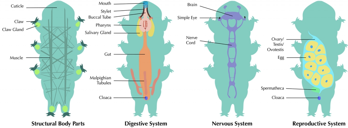
Eight Clawed Legs That Grip and Waddle
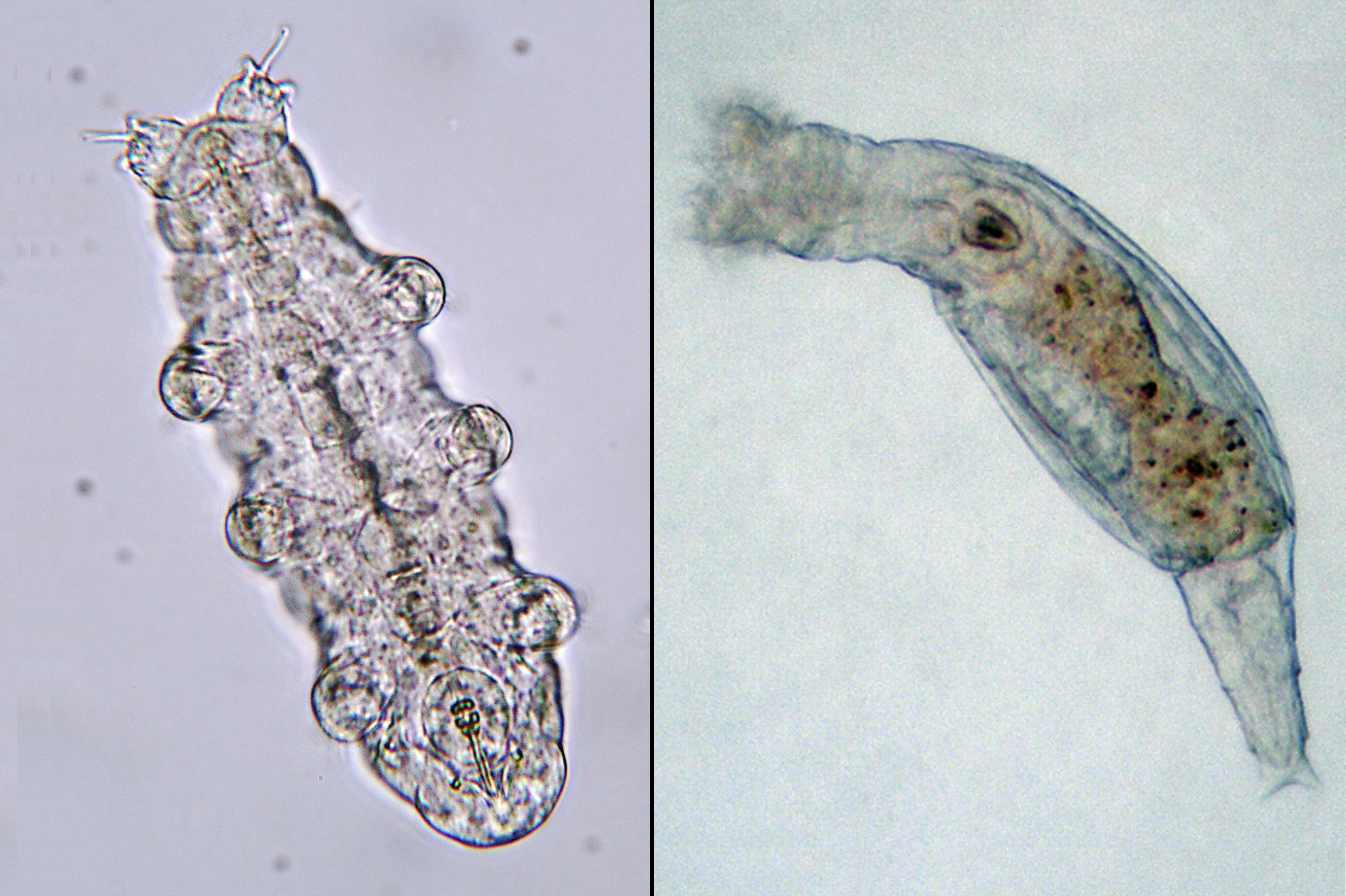
Under the microscope, the first thing you’ll notice is the eight short, segmented legs arranged in four pairs. Each leg ends in long, curved claws—usually 4 to 8 per leg—that help the tardigrade cling to moss fibers in the wild.
On a glass slide, they may appear to flail or “doggie paddle” due to lack of traction. But their natural movement is a slow, bear-like waddle, especially visible when viewed from the ventral (belly-first) angle. This unique gait is a key identifier—no other common microorganism moves quite like it.
Translucent Body Reveals Internal Organs
Tardigrades have a plump, segmented body that looks almost cartoonish under magnification. Their outer cuticle is typically translucent or milky white, allowing you to see internal structures.
Watch for:
– A pulsing pharynx near the head (sign of feeding)
– A visible gut that may be green, yellow, or brown—colors come from ingested algae or plant cells
– Occasional molting events, where the tardigrade sheds its skin, sometimes leaving behind an empty cuticle with claw tips still intact
The body contracts and expands slightly with movement, and in stressed conditions, it may begin to shrink into a tight tun—a survival ball formed during dehydration.
Mouthparts Designed for Piercing and Sucking
At the front end, you can observe the buccal apparatus, a complex feeding structure. It includes:
– Stylets: Needle-like mouthparts used to pierce plant or prey cells
– Pharyngeal pump: A muscular bulb that creates suction to draw in fluids
When feeding, the pump contracts rhythmically—look for a pulsing motion just behind the mouth. Predatory species may be seen attacking rotifers, while herbivorous ones probe moss fragments.
Best Microscope Settings for Clear Viewing
Choose 40x to 100x Total Magnification
You don’t need extreme power to see a water bear clearly:
– 40x–60x (stereo scope): Ideal for scanning and tracking movement
– 100x–200x (compound scope): Best for observing claws, mouthparts, and internal activity
Higher magnifications (400x+) reduce depth of field and can make focusing difficult. Start low, locate your specimen, then zoom in gradually.
Enhance Visibility with Advanced Lighting
Standard brightfield works, but these techniques reveal more:
Darkfield Microscopy
Creates a high-contrast image—tardigrades glow against a dark background, making leg movements and claw details pop.
Phase Contrast
Improves clarity of internal structures like muscles, nerves, and nuclei. Excellent for educational or research use.
DIC (Differential Interference Contrast)
Gives a 3D-like appearance with exceptional surface detail. Often used in scientific imaging.
Polarized Light
Highlights birefringent tissues like muscle fibers, producing stunning rainbow patterns—popular in microscopic art.
Behavior to Watch For on the Slide
Slow, Purposeful Crawling Through Water Films
Tardigrades don’t rush. Their movement is deliberate and meandering, often pausing as if “thinking.” This isn’t confusion—it’s exploration. In nature, they use their claws to crawl through microscopic water channels in moss, seeking food or mates.
On a slide, they may seem disoriented. Reduce light intensity to prevent overheating and give them time to acclimate.
Tun Formation: Witnessing Suspended Life
If the water starts to evaporate or temperature shifts, you might see tun formation:
– The body contracts into a tight, barrel-shaped ball
– Legs pull inward
– Movement stops entirely
This cryptobiotic state allows survival in extreme conditions. Add a drop of water, and within minutes, the tardigrade may unfurl and resume activity—even after years of dormancy.
Feeding and Digestion in Real Time
Active tardigrades display clear feeding behavior:
– Stylets extend and pierce food sources
– Pharyngeal pump pulses every few seconds
– Gut fills with colored fluid (often green from algae)
You might even witness defecation—a tiny release of waste from the rear. Harmless and fascinating, it confirms the animal is metabolizing food.
How to Tell Tardigrades Apart from Look-Alikes
Rotifers: Fast Spinners Without Legs
Rotifers are smaller and move quickly, often with a crown of spinning cilia that resembles a rotating wheel. They lack legs and claws and dart erratically—nothing like the slow waddle of a water bear.
Nematodes: Slithering Threads
Roundworms (nematodes) are long, thin, and smooth, moving in undulating S-shapes. They have no segmentation or limbs and glide like eels through water.
Mites: Flattened and Speedy
Microscopic mites have eight legs but are flatter, faster, and more insect-like. They don’t contract into tuns and are rarely found in moss water samples.
Key ID Tip: Only tardigrades combine eight clawed legs, barrel-shaped body, slow gait, and tun-forming ability.
Why Tardigrades Can Survive Space, Heat, and Ice
Tun State: Life Without Water or Metabolism
In harsh conditions, tardigrades enter anhydrobiosis, a form of cryptobiosis:
– Lose up to 97% of body water
– Replace fluids with protective proteins (CAHS, SAHS)
– Form a glass-like matrix inside cells to prevent damage
This lets them survive:
– Temperatures from -272°C to 150°C
– Pressures up to 6,000 atmospheres
– Space vacuum and UV radiation
In 2007, tardigrades were exposed to space for 10 days—many survived, rehydrated, and reproduced.
Dsup Protein: Nature’s DNA Shield
Tardigrades produce Dsup (Damage Suppressor) protein, which binds to DNA and protects it from radiation. Studies show it reduces X-ray damage by 40%—a discovery with potential for human medicine, including radiation therapy protection.
Are Water Bears Dangerous? No—They’re Harmless
Tardigrades are not parasitic, toxic, or invasive. They pose no threat to:
– Humans (even if ingested)
– Pets
– Plants
They are fully formed animals, not bacteria or viruses. If you drink water containing them, they’ll be digested safely.
Educational and Scientific Value of Tardigrades
Ideal for Classroom Microscopy
Tardigrades teach:
– Microscopy skills
– Cell biology
– Ecology and adaptation
Their hardiness allows long-term observation of molting, feeding, and cryptobiosis.
Citizen Science and Global Research
Amateurs contribute by:
– Posting findings on iNaturalist or TikTok with #Tardigrade
– Joining global biodiversity surveys
– Testing urban vs. wild environments
Live cultures are available from suppliers like Carolina Biological, making them accessible without field collection.
How Long Do Tardigrades Live?
Active Life: 3–4 Months (Up to 2 Years)
Most live 3 to 4 months, though some species reach 2 years. Growth happens through cell enlargement, not division—a trait called eutely.
Dormant Survival: Over 30 Years
In the tun state, they’ve survived over 30 years in labs. Theoretical models suggest century-long survival.
Fascinating Fun Facts
- Only animals to survive unprotected space exposure (FOTON-M3 mission, 2007)
- Meme nicknames: “microscopic demons,” “Captain Tardigrade, Defender of the Multiverse”
- Inspired plush toys, D&D monsters, and South Park episodes
- Some say they resemble Patrick Stewart in a spacesuit
“Finding your first tardigrade is a big deal. It’s so exciting… Everyone should experience it.” – Justine Dees, PhD
Final Tips for Finding and Observing Water Bears
- Be patient—your first find may take several tries
- Store samples in the fridge with water for weeks of reuse
- Use a phone adapter to capture photos or videos
- Share your discovery—join the global community of tardigrade hunters
Final Note: Whether you’re a student, educator, or curious hobbyist, seeing a water bear under microscope is more than a scientific observation—it’s a moment of wonder. In that tiny, waddling creature lies a story of survival, adaptation, and the incredible resilience of life. With just a slide, a drop of water, and a little patience, you can witness one of nature’s most extraordinary beings up close.


