A single drop of pond water holds a universe invisible to the naked eye—one where green spirals twist slowly, glass-like shells glide silently, and spinning cells flash red eyespots toward the light. When you place that drop under a microscope, algae emerge as the stars of the microscopic world. These tiny photosynthetic powerhouses are responsible for generating up to 50% of Earth’s oxygen, yet most people never see them until magnified. Viewing algae under the microscope transforms a murky puddle into a living gallery of nature’s engineering: from the elegant symmetry of diatom frustules to the mesmerizing spiral chloroplasts of Spirogyra.
Whether you’re a student dissecting a biology lab, a hobbyist exploring backyard ponds, or an aquarium owner battling green scum, understanding what algae look like—and why they matter—starts with observation. With just a compound microscope and a wet mount, you can identify different types, distinguish true algae from lookalike bacteria, and even detect early signs of harmful blooms. This guide walks you through exactly what to expect, how to find it, and what those tiny green threads or golden shells reveal about the health of aquatic ecosystems.
How to Spot Algae on a Microscope Slide
Look for Internal Color and Cellular Structure
The first clue that you’re looking at algae under the microscope is internal coloration. Unlike bacteria or detritus, which appear uniformly transparent or grainy, algae contain plastids—specialized organelles like chloroplasts that capture sunlight. These show up as bright green, golden, or brown patches inside the cell. If you see a cell with distinct green bands, star-shaped clusters, or cup-like structures inside, you’ve likely found algae.
Cells without internal color are probably protozoa, cyanobacteria, or non-living particles. True algae will display organized, membrane-bound organelles—especially at higher magnifications (400x). The presence of a visible nucleus further confirms a eukaryotic alga, differentiating it from prokaryotic cyanobacteria.
Pro tip: At 100x magnification, watch for cytoplasmic streaming—the slow movement of chloroplasts within the cell. This dynamic flow confirms metabolic activity and is a hallmark of living algae.
Analyze Cell Shape and Organization
Algae come in four main structural forms, each offering visual clues:
- Unicellular: Single, round or oval cells like Chlamydomonas, often zipping across the field with flagella.
- Colonial: Groups of identical cells forming geometric spheres, such as Volvox.
- Filamentous: Chains of cells resembling green threads—common in Spirogyra and aquarium hair algae.
- Flagellated: Motile cells like Euglena, moving with a whip-like flagellum.
These shapes help narrow down identification before diving into species-level details. A long, segmented green strand? Likely filamentous green algae. A slow-gliding, boat-shaped cell with golden color? Probably a diatom.
Identify Algae by Key Microscopic Features

Chloroplasts: The Signature of Photosynthesis
Chloroplasts are the definitive feature of algae. In green algae, they’re bright green due to chlorophyll a and b. But their shape is what makes them powerful ID tools:
- Spiral: Spirogyra – unmistakable helical bands wrapping around the cell
- Star-shaped: Zygnema – radiating chloroplasts with central nuclei
- Cup-shaped: Chlamydomonas – fills most of the pear-shaped cell
- Reticulate (net-like): Oedogonium – intricate green network
At 400x, you can often see chloroplasts moving—proof of active cytoplasmic streaming. If you see structured green organelles, the organism is almost certainly a eukaryotic alga, not a bacterium.
Cell Walls and Frustules: Armor and Architecture
External structures vary dramatically by group:
- Green algae: Flexible cellulose walls allow slight shape changes
- Diatoms: Rigid, glass-like silica frustules with species-specific patterns
- Dinoflagellates: Stiff cellulose “armor” plates give angular shapes
- Cyanobacteria: No true wall—just a peptidoglycan layer, appearing smooth and uniform
Diatom frustules are some of the most beautiful structures in biology. Under oil immersion (1000x), their radial or bilateral symmetry, pores, and ridges resemble microscopic stained glass.
Motility: How Does It Move?
Watch movement for 30 seconds. Is it gliding, spinning, or darting?
- Flagellar motion: Euglena moves with a snake-like wriggle; dinoflagellates spin due to a transverse flagellum
- Gliding: Some diatoms creep slowly across the slide
- Passive drift: Filamentous algae sway with water currents but don’t self-propel
If a cell moves toward light, check for an eyespot (stigma)—a red-orange dot near the flagellum in Euglena. This light-sensing organelle guides phototaxis, helping the cell find optimal light for photosynthesis.
Common Types of Algae Seen Under the Microscope
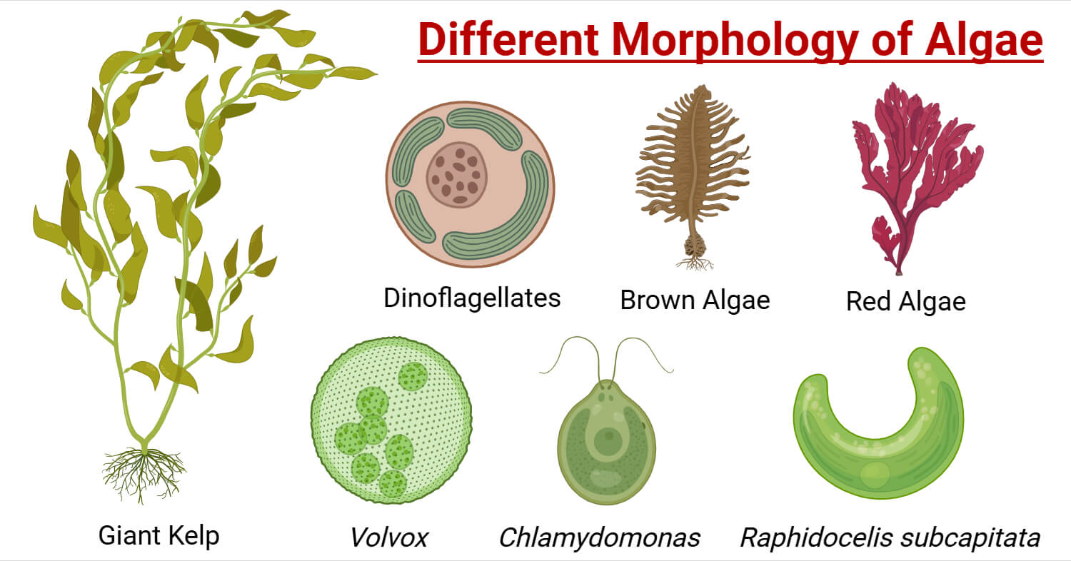
Spirogyra: The Green Spiral Alga
One of the most recognizable freshwater algae, Spirogyra forms long, unbranched filaments. At 100x–400x, its spiral chloroplasts are unmistakable—green ribbons coiling around the nucleus like DNA strands.
- Size: 10–100 µm wide, filaments up to several mm long
- Habitat: Ponds, ditches, aquariums
- Reproduction: During conjugation, filaments align and form ladder-like tubes—visible under the scope
Volvox: The Living Sphere
Volvox colonies look like tiny green snow globes, 0.5–1 mm in diameter. Each sphere contains hundreds of flagellated cells embedded in a gelatinous matrix. Inside, you may see smaller daughter colonies developing.
- Movement: Smooth, rolling motion
- Best viewed at: 100x
- Habitat: Nutrient-rich stagnant water
Diatoms: Nature’s Microscopic Artists
Diatoms are unicellular algae with silica frustules that persist long after death. Two main types:
- Radial (centric): Circular or star-shaped (Coscinodiscus)
- Pennate (bilateral): Elongated, boat-shaped (Navicula, Nitzschia)
Many pennate diatoms glide slowly across the slide—look for this subtle movement. Their golden-brown color comes from fucoxanthin, an accessory pigment.
Fun fact: Diatom patterns are so precise, they’ve inspired nanotechnology and even micro-sculptures.
Dinoflagellates: The Spinning Toxins
Common in marine samples, dinoflagellates have two flagella: one encircles the cell (causing spin), the other trails behind. Many have angular, armored plates.
- Bioluminescence: Some species flash light when disturbed
- Toxic species: Alexandrium and Gonyaulax produce saxitoxins, causing paralytic shellfish poisoning
Watch for rapid spinning motion—this is a key identifier.
Cyanobacteria: Not True Algae
Despite the nickname “blue-green algae,” cyanobacteria are prokaryotes. They lack nuclei and chloroplasts, appearing as uniform blue-green filaments or colonies.
- Filamentous types: Anabaena (with round heterocysts for nitrogen fixation), Oscillatoria (smooth, wavy strands)
- Colonial: Microcystis forms slimy green rafts—often toxic
Warning: Microcystis blooms produce microcystins, which can damage the liver.
Euglena: The Mixotrophic Survivor
Euglena looks like a green, elongated cell with a faint red eyespot and one hard-to-see flagellum. Without a rigid cell wall, it can change shape.
- Size: 15–40 µm
- Movement: Wriggling, snake-like motion
- Unique trait: Can switch to eating organic matter in the dark (mixotrophy)
Common in nutrient-rich, stagnant water.
Filamentous Algae in Ponds and Aquariums

Green Hair Algae
Long, tangled green threads in aquariums are usually green filamentous algae like Spirogyra or Oedogonium. Microscopically:
- Rectangular cells in chains
- Visible green chloroplasts
- No branching
Cause: High nitrates, excess light, low CO₂
Brown Hair Algae (BHA)
Despite the name, BHA is not green algae—it’s a diatom, often Vaucheria. Appears as:
- Short, fuzzy brown growth on glass or plants
- Golden-brown plastids due to fucoxanthin
- Cells show segmentation and nuclei at high power
Usually appears in new tanks and fades as silicates deplete.
Green Tuft Algae
Forms dense, spherical clumps of fine filaments. Often mistaken for Cladophora.
- Branched or radiating structure
- High surface area for light capture
- Indicates high nutrients and moderate light
How to Prepare a Wet Mount for Algae Observation
Step-by-Step Slide Preparation
- Collect sample: Use a pipette to draw water from pond, aquarium, or bloom.
- Place drop on slide: One small drop prevents overflow.
- Add coverslip: Lower at 45° to avoid bubbles.
- Blot excess: Use paper towel to wick away liquid.
- Start at 40x: Scan for larger structures, then increase to 100x or 400x.
Pro tip: Add a speck of methyl cellulose to slow fast-moving organisms like Euglena.
Best Microscopes for Viewing Algae
Compound Microscope (40x–1000x)
Essential for detail:
– 40x: View whole filaments or colonies
– 400x: See chloroplasts, nuclei, flagella
– 1000x (oil immersion): Diatom frustule patterns
Recommended: AmScope M150C-I, Omax 40X-2000X, Nikon Eclipse E100
Dissecting Microscope (10x–50x)
Useful for observing motility and isolating specimens. Pair with compound scope for full analysis.
Algae in the Microbial Food Web
Algae feed a rich microfauna:
– Daphnia: Transparent bodies with green guts from eaten algae
– Rotifers: “Wheel animals” with ciliated coronas that suck in algae
– Copepods: Jerky swimmers that filter-feed on phytoplankton
– Paramecium: Ciliated protists with green food vacuoles
Note: Water mites are predators—they hunt micro-crustaceans, not algae.
What Algae Reveal About Water Quality
| Algae Type | Water Condition |
|---|---|
| Green filamentous | High nutrients, excess light |
| Diatoms (brown) | Low silicates, early tank phase |
| Cyanobacteria | Eutrophication, low oxygen |
| Dinoflagellates | Warm, nutrient-imbalanced marine systems |
Harmful blooms show dense Microcystis colonies or Anabaena filaments with heterocysts—early warning signs for environmental monitoring.
Final Tips for Microscopic Exploration
- Start with green algae and diatoms
- Use multiple magnifications
- Let samples settle for clearer motion
- Sketch or photograph findings
- Share with communities like r/Microscopy or tag @joyfulmicrobe
Observing algae under the microscope isn’t just science—it’s a window into the invisible forces sustaining life. With a drop of water and a lens, you’re not just looking at algae. You’re seeing the pulse of the planet.