
Finding a tiny speck crawling across your sheets can send your mind racing—could it be a bed bug? While adult bed bugs are visible to the naked eye, examining a specimen under a microscope is often the only way to confirm an infestation with certainty. At high magnification, what appears as a simple brown oval transforms into a complex, highly evolved parasite built for stealth and survival.
The bed bug under microscope reveals a world of specialized anatomy: piercing mouthparts designed for blood-feeding, sensory bristles that detect your slightest movement, and a flattened body shaped to vanish into the tiniest cracks. This level of detail isn’t just fascinating—it’s essential for accurate identification, especially when dealing with early-stage nymphs or look-alike pests.
Whether you’re troubleshooting unexplained bites, inspecting secondhand furniture, or simply curious about what’s hiding in your home, understanding how bed bugs appear under magnification empowers you to act confidently. This guide explores the microscopic features of Cimex lectularius, compares life stages, distinguishes real threats from false alarms, and shows how science confirms what’s truly lurking in your bedroom.
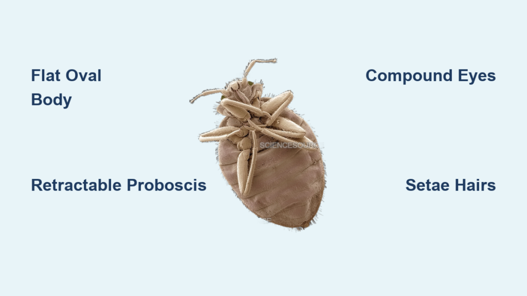
Flat, Oval Body with Segmented Abdomen
At low to medium magnification (10x–40x), the bed bug’s dorsoventrally flattened body becomes clearly segmented into three main parts: head, thorax, and abdomen. The exoskeleton is rigid and composed of chitin, giving it a smooth yet textured appearance under higher power.
The 11 abdominal segments are ringed with fine ridges and covered in microscopic setae—small bristle-like projections that give the surface a slightly fuzzy or velvety look. This flattened shape allows bed bugs to squeeze into crevices as narrow as 0.5 mm, such as mattress seams, baseboard gaps, or electrical outlet covers.
Unlike many insects, bed bugs lack wings entirely. Their streamlined, wingless form is optimized for concealment, not flight. Under the microscope, this adaptation becomes obvious—every feature supports a life spent hidden in darkness, emerging only to feed.
Retractable Proboscis and Piercing Mouthparts
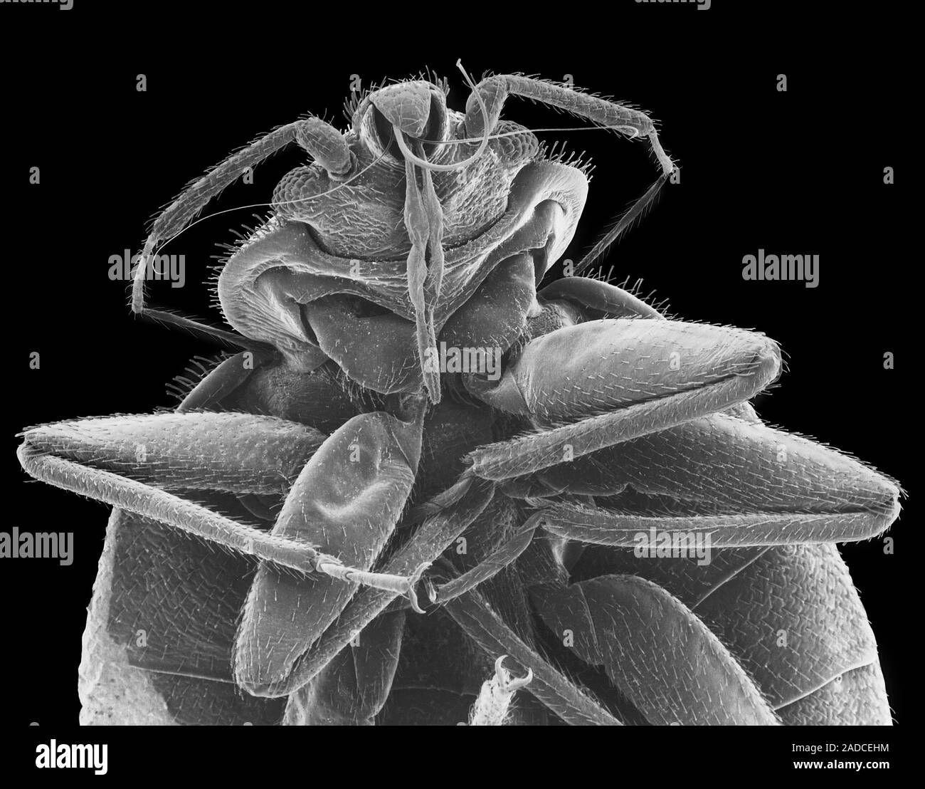
One of the most definitive signs of a bed bug under microscope is the presence of a needlelike proboscis tucked beneath the head. At rest, this feeding tube lies folded under the body, but under 40x magnification or higher—especially with scanning electron microscopy (SEM)—its segmented, jointed structure becomes clear.
During feeding, the proboscis extends forward and pierces the skin using two sharp stylets:
– One injects anticoagulant saliva to prevent blood from clotting.
– The other draws blood directly into the digestive tract.
These are not chewing mouthparts. Instead, they’re precision tools for piercing and sucking, confirming the insect’s hematophagous (blood-feeding) nature. In color-enhanced micrographs from institutions like the CDC, the proboscis is often highlighted in purple or red to emphasize its function.
If you see a proboscis and compound eyes under magnification, you’re almost certainly looking at a bed bug.
Prominent Compound Eyes on Head Sides
Even at 20x magnification, the compound eyes of a bed bug stand out. Positioned on either side of the head, each eye is made up of multiple ommatidia units sensitive to motion and changes in light. This visual system helps the insect detect host movement, avoid sudden light exposure, and navigate toward warmth and carbon dioxide—key cues for finding a blood meal.
Unlike some similar pests like booklice, bed bugs have large, well-developed eyes. This trait supports their nocturnal behavior and is easily confirmed under magnification, serving as a quick differentiator from non-biting insects.
Setae: Sensory Hairs Across the Body
Covering the dorsal and lateral surfaces are dozens of tiny chitinous bristles called setae. These aren’t hairs in the traditional sense but rigid projections extending from the cuticle. Under 100x magnification, they appear like small spikes or spines.
These structures act as mechanoreceptors, detecting:
– Air currents from human movement.
– Vibrations from footsteps or shifting bedding.
– Physical contact while crawling through tight spaces.
Their density gives nymphs and adults a slightly “fuzzy” appearance when viewed from above—especially under strong lighting. This texture helps distinguish bed bugs from smoother-bodied insects like mites or young cockroaches.
First Instar Nymphs: Nearly Invisible Without Magnification

Newly hatched nymphs measure only 1 to 1.5 mm—about the size of a poppy seed—and appear translucent or pale white immediately after emerging. Under the microscope, their bodies may seem almost glassy, with internal structures faintly visible.
Key identifiers:
– No red coloration until after their first blood meal.
– Abdomen expands and darkens (to red or black) after feeding.
– Eyes and setae are present but less developed than in later stages.
Because they lack the rusty-brown hue of adults, first instar nymphs are frequently missed during visual inspections. Only microscopic examination can reliably confirm their presence—critical for early detection before populations explode.
Later Instars (2nd–5th): Gradual Darkening and Growth
With each molt, nymphs increase in size and pigmentation:
– Second instar: ~2 mm, light tan.
– Third instar: ~3 mm, darker tan.
– Fourth and fifth instars: 4–5 mm, increasingly brown.
Microscopy shows progressive development of:
– Eye complexity.
– Density of setae.
– Sharpness and strength of mouthparts.
They remain smaller and paler than adults, but their increasing size makes them more detectable even without magnification—especially after feeding, when the abdomen swells and darkens.
Adult Bed Bugs: Full Size and Distinct Coloration

Adults reach 5 to 7 mm in length—still small, but clearly visible without tools. Under the microscope:
– Rusty brown to reddish-brown exoskeleton.
– Engorged abdomen appears deep red or purple-black after feeding.
– Clearly defined segmentation and dense setae coverage.
High magnification reveals the pronounced thoracic structure and well-developed legs, each ending in small claws adapted for gripping fabric and wood. Males may show subtle differences in abdominal tip morphology related to traumatic insemination, though this typically requires dissection or extreme magnification.
Scanning Electron Microscopy (SEM): 3D Surface Detail
Scanning electron microscopy provides ultra-high-resolution, three-dimensional images of bed bug surfaces. This technique is used in scientific studies to examine:
– Fine texture of the exoskeleton.
– Structure of individual setae.
– Mechanics of mouthpart deployment.
Because SEM produces grayscale images, researchers often digitally colorize features (e.g., red eyes, purple proboscis) to enhance clarity. These visuals are widely shared by the CDC and Harvard University, helping professionals and the public recognize key traits.
Light Microscopy: Practical Field Identification
For non-laboratory settings, light microscopy (compound or stereo microscopes) is sufficient for identifying live or preserved specimens. At 10x to 100x magnification:
– Movement patterns can be observed.
– Fecal droppings appear as dark, melanized specks.
– Eggs (1 mm, pearly white) can be examined for viability.
Handheld digital microscopes (available to consumers) offer real-time imaging and are useful for inspecting mattress seams, headboards, or suspicious spots.
Scale References in Micrographs
Many reference images include graph paper backgrounds with 1/4 inch squares to illustrate scale. For example:
– An adult female bed bug spans nearly half a square.
– A first instar nymph fits within a corner of one square.
These comparisons help contextualize size differences between nymphs and adults—and why early-stage infestations go unnoticed.
Booklice vs Bed Bugs: Critical Microscopic Differences
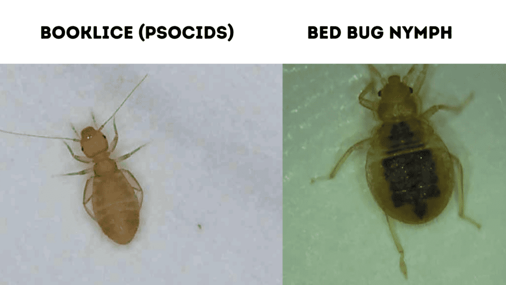
A common misidentification occurs between bed bugs and booklice (psocids), both of which are small, pale, and found in homes. But under magnification, they are easily distinguished.
| Feature | Bed Bug | Booklouse |
|---|---|---|
| Size | 1–7 mm | 1–2 mm |
| Color | Brown, reddish after feeding | Translucent, grayish |
| Body Shape | Flat, oval, hard exoskeleton | Soft, elongated, no segmentation |
| Eyes | Large compound eyes | Tiny or absent eyes |
| Mouthparts | Proboscis (piercing-sucking) | Chewing mouthparts |
| Setae | Dense, stiff bristles | Sparse, fine hairs |
| Feeding Evidence | Blood-filled abdomen, fecal spots | Found near mold or damp paper |
✅ Definitive confirmation: Presence of compound eyes + proboscis + blood residue = bed bug.
Booklice do not bite humans and feed on fungi, mold, and starches—commonly found in damp books or wallpaper glue. If your “bed bug” lacks piercing mouthparts and has a soft body, it’s likely a harmless psocid.
Confirming Hidden Infestations
In one documented case, a person experienced unexplained hives for months despite thorough cleaning. No bugs, droppings, or shed skins were found—until a tiny insect was captured and examined under a digital microscope.
Analysis revealed:
– Pale, translucent body.
– Size of ~1.2 mm.
– Clear segmentation, compound eyes, and retractable proboscis.
Entomologists confirmed it as a first instar nymph, indicating a recent hatch or low-level infestation. The absence of visible signs explained the delayed discovery—highlighting how microscopy enables early intervention.
Why Only One Person Gets Bitten
Interestingly, only one partner showed bites, despite sharing the bed. This is common: human reactions to bed bug saliva vary widely. Some develop large, itchy welts; others show no reaction at all. Microscopic confirmation bypasses reliance on bite patterns, offering objective evidence of presence regardless of symptoms.
Using Magnification for Early Detection
You don’t need a lab microscope to spot early signs. Handheld digital microscopes ($30–$100) connect to smartphones or computers and allow detailed inspection of:
– Mattress seams and stitching.
– Box spring joints.
– Headboard crevices.
– Behind wall outlets near the bed.
Look for:
– Live bugs, especially pale ones (nymphs).
– Fecal specks: dark, ink-like stains that smear when wet.
– Eggs: pearly white, ~1 mm, often laid in clusters.
– Shed exoskeletons (exuviae): golden-brown, hollow shells left after molting.
Magnification turns subtle clues into clear evidence.
Why Alcohol Doesn’t Work
Despite viral DIY advice, rubbing alcohol kills only up to 50% of bed bugs on direct contact and does not penetrate eggs. Rutgers University studies show it fails to eliminate hidden populations.
More critically:
– Highly flammable, posing fire risks when sprayed on mattresses or furniture.
– Not residual, meaning bugs returning after treatment survive.
⚠️ Official warning (NJAES): “Rubbing alcohol should not be used to control bed bugs.”
Source: https://njaes.rutgers.edu/fs1251/
Microscopy shows why: alcohol cannot reach bugs deep in cracks or affect eggs sealed in crevices.
Proven Control Methods
Heat Treatment: Kills All Life Stages
Temperatures above 118°F (48°C) sustained for 90+ minutes kill adults, nymphs, and eggs. Heat penetrates fabric, wood, and upholstery—reaching where sprays and alcohol cannot.
Steam Cleaning: Immediate Kill on Contact
Handheld steamers (150°F+ at nozzle tip) kill visible bugs and eggs in seams, tufts, and baseboards. Use slowly and methodically—microscopic eggs are tiny and easily missed.
Mattress Encasements: Trap and Monitor
Allergen-proof covers prevent bugs from entering or escaping. Trapped insects will eventually die. Use with regular inspection (aided by magnification) to monitor success.
Final Takeaways: Why Microscopy Matters
Examining a bed bug under microscope transforms uncertainty into clarity. It reveals:
– Anatomical proof of blood-feeding adaptations.
– Developmental stages that explain elusive infestations.
– Critical differences from harmless insects like booklice.
– Limits of DIY remedies and need for professional strategies.
🔍 Bottom line: If you find a tiny bug in your bed, verify it under magnification before panicking—or ignoring it. The difference between a bed bug and a booklouse could mean the difference between professional treatment and unnecessary fumigation.


