
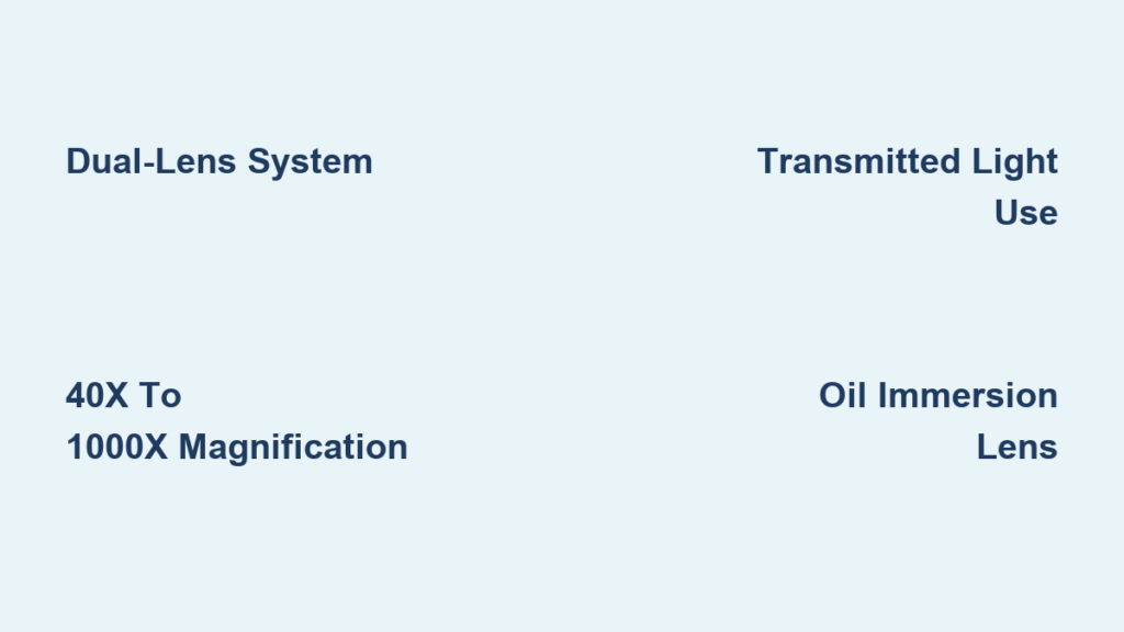
If you’ve ever observed a cheek cell, a strand of onion skin, or a single-celled amoeba under a microscope, you’ve likely used a compound microscope. This powerful optical instrument is designed to magnify tiny, transparent specimens using a dual-lens system—delivering clear, highly detailed images of structures far too small to be seen with the naked eye. The compound microscope definition centers on its use of two or more lenses—an objective lens and an eyepiece (ocular) lens—that work together to achieve magnification from 40x to 1000x, making it indispensable in biology, medicine, and education. Unlike a simple microscope, which uses just one lens, the compound microscope produces a virtual, inverted, and two-dimensional image by focusing light through the specimen. In this comprehensive guide, you’ll learn the full compound microscope definition, how it functions, its key components, and why it remains a vital tool across scientific disciplines.
What Makes a Microscope “Compound”?
The term “compound” refers to the use of multiple lenses in sequence to magnify an image. This is the core of the compound microscope definition. While a simple microscope, like a magnifying glass, uses a single lens, a compound microscope combines at least two converging lenses: the objective lens, positioned close to the specimen, and the eyepiece lens, which the viewer looks through. Light passes through the specimen from below, through the objective lens, and then through the eyepiece, resulting in a highly magnified, inverted image.
Because it relies on transmitted light—light passing through the specimen—it is also known as a bright-field or transmitted light microscope. This setup works best with thin, transparent samples mounted on glass slides. The dual-lens system allows for significantly higher magnification and resolution than single-lens instruments, enabling scientists and students to study cells, bacteria, and internal tissue structures with remarkable clarity.
Key Optical Components Explained

Objective Lenses: The Primary Magnifiers
Mounted on a rotating nosepiece, the objective lenses are responsible for the first stage of magnification. Most compound microscopes come equipped with four standard objectives:
- 4x (scanning) – Offers a broad field of view for initial specimen location
- 10x (low power) – Provides a general overview of larger structures
- 40x (high power) – Delivers detailed observation of cellular features
- 100x (oil immersion) – Achieves maximum resolution and is used with immersion oil
Each objective lens forms a real, inverted image inside the body tube. The 100x oil immersion lens is unique: by placing a drop of immersion oil between the lens and the slide, it minimizes light refraction and increases resolution. The oil’s refractive index closely matches that of glass, allowing more light to enter the lens and reducing image distortion.
Eyepiece (Ocular Lens): Final Image Enlargement
The eyepiece, or ocular lens, further magnifies the image created by the objective. Most standard eyepieces are 10x, though 5x, 15x, and 20x options exist. Binocular microscopes include prisms that split the image so both eyes receive identical views, reducing eye strain during extended use.
Total magnification is calculated using the formula:
Objective Magnification × Eyepiece Magnification
For example:
– 10x objective × 10x eyepiece = 100x total magnification
– 100x oil immersion objective × 10x eyepiece = 1000x total magnification
This multiplicative effect is what enables the compound microscope to reveal microscopic details invisible to the human eye.
Condenser and Diaphragm: Precision Light Control
Located beneath the stage, the condenser focuses light onto the specimen. Adjusting its height ensures optimal illumination and image clarity. Below the condenser is the iris diaphragm, which controls the diameter of the light beam. Closing it slightly increases contrast by reducing scattered light, while opening it enhances brightness—though excessive opening can wash out fine details.
Illuminator: Consistent Light Source
Modern compound microscopes feature built-in LED or halogen illuminators at the base. These provide bright, adjustable, and consistent light without relying on external sources. Older models used mirrors (plane for bright light, concave for dim conditions), but modern electric lighting ensures stable, uniform illumination, crucial for accurate observation.
Mechanical Parts and Their Functions
Base and Arm: Structural Stability
The base (or foot) is a heavy, U-shaped metal support that prevents the microscope from tipping. The arm extends upward from the base and is used to carry the microscope. It supports the stage, body tube, and focusing mechanisms.
Stage and Mechanical Stage: Specimen Handling
The stage is a flat platform with a central opening for light to pass through. It holds the slide in place using stage clips. Advanced models feature a mechanical stage with precision knobs that allow controlled movement of the slide in the X and Y directions—essential for scanning large specimens without losing focus.
Body Tube and Draw Tube: Optical Alignment
The body tube maintains the correct distance between the objective and eyepiece lenses, ensuring proper focus. The draw tube at the top holds the eyepiece and may be adjustable in some models for fine-tuning focus.
Focusing Mechanisms: Coarse and Fine Knobs
- Coarse adjustment knob: Moves the stage (or body tube) rapidly for initial focusing, ideal for low magnification (4x and 10x).
- Fine adjustment knob: Allows minute movements for sharp focusing at high magnification (40x and 100x).
- Automatic stop: Prevents the objective lens from crashing into the slide during coarse focusing—protecting both the lens and specimen.
Inclination Joint: Ergonomic Viewing
Some models include an inclination joint, allowing the head to tilt for comfortable viewing while seated. However, tilting is not recommended when viewing liquid specimens, as they may slide off the cover slip.
How Magnification and Resolution Work

Total Magnification Range
Standard compound microscopes offer magnifications from 40x to 1000x:
| Objective | Eyepiece | Total Magnification |
|---|---|---|
| 4x | 10x | 40x (scanning) |
| 10x | 10x | 100x (low power) |
| 40x | 10x | 400x (high power) |
| 100x | 10x | 1000x (oil immersion) |
Beyond 1000x, empty magnification occurs—images appear larger but no additional detail is visible due to the physical limits of visible light.
Maximum Useful Magnification
The theoretical maximum is about 2000x, beyond which resolution does not improve. This limit is imposed by the wavelength of visible light (400–700 nm), which restricts how finely details can be distinguished.
Resolution: Clarity Beyond Magnification
Resolution—the ability to distinguish two closely spaced points as separate—is more important than magnification. The compound microscope’s resolution limit is approximately 200 nanometers, based on Abbe’s diffraction limit. This means it can clearly resolve bacteria (0.2–2 μm) and cell nuclei (~5–10 μm), but not smaller structures like viruses (<200 nm), which require electron microscopes.
Specialized Types of Compound Microscopes
Phase Contrast Microscope
Enhances contrast in transparent, unstained specimens like live cells and bacteria by converting phase shifts in light into brightness differences. Uses a phase contrast objective and a phase slider in the condenser.
Best for: Cell biology, microbiology, live-cell imaging.
Polarizing Microscope
Uses a polarizer and analyzer to cross-polarize light, revealing birefringent materials that split light into two rays.
Best for: Mineral analysis, crystal structure studies, pharmaceuticals.
Metallurgical Microscope
Designed for opaque specimens like metals and ceramics. Uses reflected light (epi-illumination) that shines down through the objective.
Best for: Industrial inspection, quality control, gemology.
Fluorescence Microscope
Excites fluorophores in the specimen with specific wavelengths (often UV or blue light). The emitted light creates bright, colored images against a dark background.
Key components: Excitation filter, dichroic mirror, emission filter.
Best for: Molecular biology, immunology, DNA/protein tagging.
Differential Interference Contrast (DIC) Microscope
Uses polarized light and interference to create a pseudo-3D image with shadow-like contrast. Reveals fine surface details in transparent specimens.
Best for: High-resolution imaging of subcellular structures.
Compound vs. Stereo Microscope: Key Differences
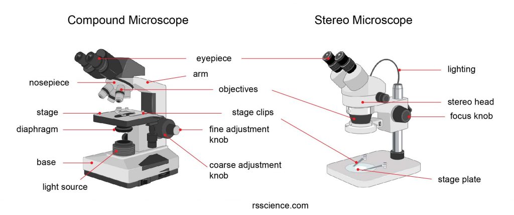
| Feature | Compound Microscope | Stereo Microscope |
|---|---|---|
| Magnification | 40x – 1000x | 10x – 100x |
| Image Type | 2D, inverted | 3D, upright |
| Light Source | Transmitted (from below) | Reflected (from above) |
| Specimen | Thin, transparent slides | Thick, opaque objects |
| Slide Required | Yes | No |
| Depth of Field | Low | High |
| Primary Use | Cellular study | Dissection, inspection |
Stereo microscopes are ideal for manipulating objects, while compound microscopes excel at revealing internal cellular details.
Advantages and Limitations
Why Use a Compound Microscope?
- High magnification and resolution for cellular-level detail
- Built-in illumination ensures consistent, bright images
- Widely used in education and labs—easy to operate and maintain
- Cost-effective compared to electron or confocal microscopes
- Versatile with multiple specialized variants available
Limitations to Consider
- Limited to 1000x magnification—cannot see viruses or molecular structures
- Requires thin, transparent specimens—opaque samples won’t transmit light
- Image is inverted and reversed—can confuse beginners
- Oil immersion lenses need special care—oil must be cleaned after use
- Low depth of field—only a thin layer of the specimen stays in focus
Common Applications Across Fields
Biology and Education
Used in schools and universities to study plant and animal cells, bacteria, protozoa, and cell division. Students learn staining techniques (e.g., methylene blue, iodine) to enhance visibility.
Medical Diagnostics
- Pathology: Examine tissue biopsies for cancer
- Hematology: Count and identify blood cells
- Microbiology: Identify bacteria and parasites in samples
Forensic Science
Analyze hair, fibers, fingerprint residue, and gunshot particles. Even minute trace evidence can be critical in criminal investigations.
Pharmaceuticals and Materials Science
- Crystal structure analysis using polarized light
- Quality control of drug formulations
- Surface inspection of metals, ceramics, and semiconductors
Environmental Science
Study plankton, algae, and waterborne pathogens in ecological and public health research.
Frequently Asked Questions
What defines a compound microscope?
A compound microscope uses two or more lenses—an objective and an eyepiece—to magnify small, transparent specimens with transmitted light. It produces a high-magnification, 2D, inverted image, distinguishing it from simple or stereo microscopes.
Why is the image inverted?
Because light rays cross as they pass through the objective lens, the image is both inverted (upside down) and reversed (left to right). This is normal and expected in compound optics.
What is the field of view?
The field of view is the diameter of the area visible through the eyepiece. It decreases as magnification increases. At 40x, you might see 4–5 mm; at 1000x, less than 0.2 mm.
What is depth of field?
Depth of field is the thickness of the specimen that remains in focus at one time. It’s inversely proportional to magnification—higher power means only a thin slice is sharp.
What is focal distance?
Also called working distance, it’s the space between the objective lens and the slide when in focus. Higher magnification objectives (like 100x) have very short working distances—sometimes less than 0.2 mm.
Are compound microscopes zoomable?
Most are fixed power, meaning you switch between discrete objectives (4x, 10x, etc.). True zooming is rare and more common in stereo microscopes.
The compound microscope remains a fundamental tool in science—not because it’s the most advanced, but because it’s reliable, accessible, and powerful for its purpose. From classrooms to research labs, it unlocks the invisible world of cells and microorganisms with clarity and precision. While electron microscopes reveal even smaller structures, the compound microscope strikes the perfect balance between magnification, resolution, and practicality. Understanding its definition, components, and capabilities empowers students, educators, and professionals to explore the microscopic universe with confidence.


