
Imagine watching a single cell divide in real time—its nucleus splitting, cytoplasm flowing, organelles shifting—all without killing or staining it. This is exactly what phase contrast microscopy makes possible. Unlike standard brightfield microscopes that rely on light absorption to create contrast, phase contrast reveals intricate details of living, unstained cells by converting invisible phase shifts in light into visible brightness differences. This technique is indispensable in biology labs for studying dynamic processes like mitosis, motility, and microbial behavior in their natural state.
At the heart of phase contrast is a clever manipulation of light waves. Transparent biological specimens—like cells and microorganisms—don’t absorb much light, but they do slow it down slightly due to differences in thickness and refractive index. These subtle delays, called phase shifts, are invisible to the human eye. However, a phase contrast microscope detects these shifts by separating light into two paths: one that passes through the specimen undisturbed (undeviated), and another that gets scattered by cellular structures (diffracted). By modifying the undeviated light and allowing it to interfere with the diffracted light, the microscope transforms phase differences into visible contrast.
In this guide, you’ll learn how phase contrast microscopy works, from the physics of light interference to the precise roles of the annular diaphragm and phase plate. You’ll discover how to align the system for optimal results, understand the difference between positive and negative phase contrast, and recognize why this method remains a cornerstone of live-cell imaging across research, diagnostics, and education.
Core Principle: Converting Phase Shifts into Visible Contrast
Transparent specimens are known as phase objects because they alter the timing (phase) of light waves rather than their intensity. Since conventional microscopes only detect amplitude (brightness), unstained cells appear nearly invisible under brightfield illumination. Phase contrast overcomes this limitation by converting phase differences into amplitude changes through controlled interference.
Light Wave Interference in Action
When light passes through a cell:
– Undeviated (direct) light travels straight through the surrounding medium.
– Diffracted (scattered) light bends around internal structures like nuclei or mitochondria and is delayed by approximately λ/4 (one-quarter wavelength) due to higher refractive index.
These two beams recombine at the image plane. Normally, their phase difference would remain undetectable. But in phase contrast microscopy, the undeviated light is manipulated so that the total phase shift between the two beams becomes λ/2 (half a wavelength), leading to destructive interference—a reduction in brightness that your eyes can see.
How Interference Creates Image Contrast
In positive phase contrast, the phase plate advances the undeviated light by an additional λ/4. Combined with the specimen-induced λ/4 delay in diffracted light, the total difference reaches λ/2. When these out-of-phase waves meet, they cancel each other out, resulting in darker regions where cellular structures are present.
This means dense organelles—like the nucleus or mitochondria—appear dark against a bright background, making them clearly visible even in living, unstained cells. The entire process hinges on precise optical engineering: transforming an invisible physical property (phase shift) into a visible visual cue (contrast).
Key Components That Enable Phase Contrast
The magic of phase contrast lies in two specially designed optical components: the condenser annulus and the phase plate. Together, they control how light interacts with the specimen to generate contrast.
Annular Diaphragm: Creating a Hollow Cone of Light
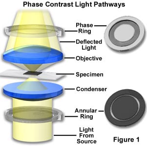
Located in the front focal plane of the condenser, the annular diaphragm is a ring-shaped aperture that shapes the illumination into a hollow cone of light. This ensures that undeviated light forms a ring-shaped wavefront when it reaches the objective’s back focal plane—perfectly matching the geometry of the phase ring.
Why it matters: Only undeviated light focuses into a ring at this plane, allowing selective manipulation by the phase plate while diffracted light spreads out and bypasses it.
Phase Plate: The Heart of Contrast Generation
Sitting in the back focal plane of the objective lens, the phase plate contains a phase ring that performs two critical functions:
1. It shifts the phase of undeviated light by λ/4 (either advancing or retarding it).
2. It reduces the amplitude of undeviated light by 60–90% using a neutral density coating.
Diffracted light, which spreads widely, mostly avoids the phase ring and remains unchanged. This selective treatment sets up the conditions for interference at the image plane.
Critical alignment: The phase ring must align perfectly with the image of the condenser annulus. Misalignment leads to poor contrast, uneven backgrounds, or ghosting.
Matching Ph1, Ph2, and Ph3 Components
Phase contrast objectives are labeled Ph1, Ph2, Ph3 based on magnification and numerical aperture:
– Ph1: Used with 10x objectives
– Ph2: Matches 20x–40x objectives
– Ph3: Designed for 60x–100x objectives
Each requires a corresponding condenser annulus. Using mismatched components—such as a Ph2 annulus with a Ph1 objective—disrupts alignment and ruins image quality.
Step-by-Step Light Path Through the Microscope
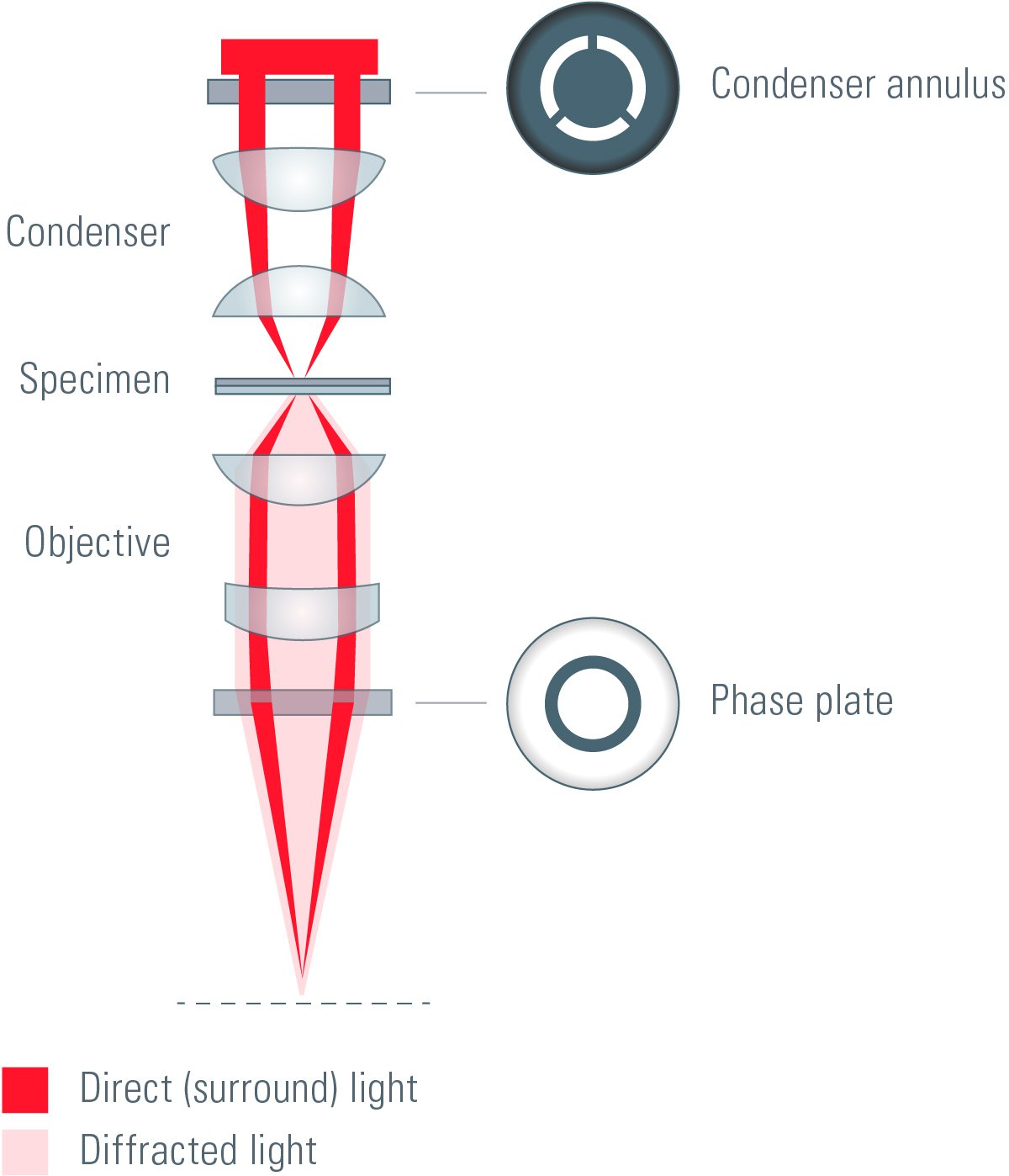
Understanding the journey of light reveals why alignment is so crucial.
1. Illumination Through the Annulus
Light from the source (LED or halogen) passes through the annular diaphragm, forming a hollow cone that illuminates the specimen uniformly.
2. Specimen Interaction
- Undeviated light passes through clear areas of the sample, maintaining its ring shape.
- Diffracted light is scattered by cellular structures, delayed by ~λ/4, and spreads over a wider angle.
3. Objective Collection
- Undeviated light converges into a ring at the objective’s back focal plane—exactly where the phase ring is located.
- Diffracted light spreads broadly and bypasses the phase ring, remaining unaltered.
4. Phase Plate Modifies Undeviated Light
The phase ring:
– Advances undeviated light by λ/4 (in positive phase)
– Dims it significantly
Now, undeviated light is λ/2 out of phase with diffracted light.
5. Interference Forms the Final Image
At the image plane:
– The two light types recombine.
– Destructive interference reduces brightness in areas with cellular structures.
– Result: Dark details on a bright background—clear visibility of nuclei, granules, and membranes.
Visual cue: Under positive phase contrast, a human cheek cell nucleus appears sharply dark, while the cytoplasm shows granular texture.
Positive vs Negative Phase Contrast: Choosing the Right Mode
Two modes offer different visual outcomes depending on your sample.
Positive Phase Contrast
- Phase plate advances undeviated light by λ/4.
- Total phase difference = λ/2 → destructive interference.
- Structures appear darker on a bright background.
- Best for: Most biological samples—cell cultures, bacteria, protozoa.
Example: Mitochondria in live fibroblasts appear as fine dark granules.
Negative Phase Contrast
- Phase plate retards undeviated light by λ/4.
- Combined with specimen-induced λ/4 delay → total = λ/2 → constructive interference.
- Structures appear brighter on a dark background.
- Best for: Protists like Vorticella, where bright cilia and nuclei stand out.
Pro tip: Use negative phase to highlight fast-moving ciliates in aquatic samples.
How to Align a Phase Contrast Microscope
Proper alignment is essential. Misalignment causes uneven backgrounds, low contrast, or ghost rings.
Step-by-Step Alignment Procedure
-
Set Köhler illumination
Focus the condenser and adjust the field diaphragm for even lighting. -
Insert phase contrast condenser
Rotate turret to match objective (e.g., Ph1 for 10x). -
Use a phase telescope or Bertrand lens
Remove an eyepiece, insert the telescope, and focus on the back focal plane. -
Align annulus with phase ring
Use condenser centering screws to superimpose the bright annulus ring over the dark phase ring. -
Repeat for each objective
Switch magnifications and realign as needed. -
Replace telescope and observe
Return eyepiece, place specimen, and adjust focus.
Warning: A misaligned system produces crescent-shaped halos or patchy brightness—always verify alignment before imaging.
Advantages of Phase Contrast Imaging
Non-Destructive Live Cell Observation
No fixation or staining required. Cells remain viable for long-term time-lapse studies of mitosis, motility, or phagocytosis.
High Contrast Without Dyes
Reveals organelles and membranes clearly in unstained cultures.
Real-Time Dynamic Monitoring
Watch cytoplasmic streaming, flagellar movement, or amoeboid motion in real time.
Compatible with Plastic Dishes
Works well with plastic petri dishes—no birefringence issues.
Cost-Effective Upgrade
Add phase contrast to existing scopes for far less than fluorescence systems.
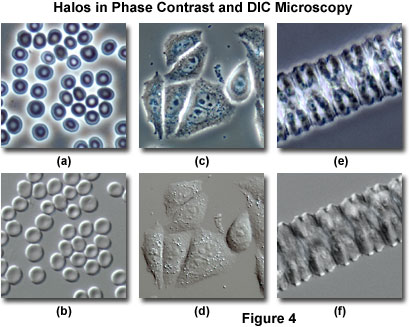
Limitations and How to Fix Them
Halo Effect
- Cause: Low-angle diffracted light interacts with phase ring.
- Solution: Use apodized phase objectives or digital filters.
Shade-Off Effect
- Cause: Uniform areas lack diffraction → no interference.
- Fix: Use higher NA objectives or switch to DIC.
Contrast Inversion
- Cause: Phase shift exceeds λ/4 → constructive interference.
- Occurs in: Thick fungal spores.
- Fix: Use negative phase or alternative method.
Reduced Resolution
- Phase annuli limit NA → slight resolution loss.
- Workaround: Use oil immersion with high-NA objectives.
Thickness Limitations
Best for thin, monolayer samples. Thick tissues cause overlapping shifts → blurred images.
Final Note: Phase contrast microscopy remains a cornerstone of biological imaging because it reveals the invisible—live, transparent cells—in stunning detail. By harnessing the wave nature of light and precise optical engineering, it turns phase shifts into contrast without harming specimens. While it has limitations like halos and thickness constraints, proper setup and understanding make it indispensable in research, diagnostics, and education. Whether you’re watching a cell divide or identifying a microbe in pond water, phase contrast brings the unseen world into clear view.


