
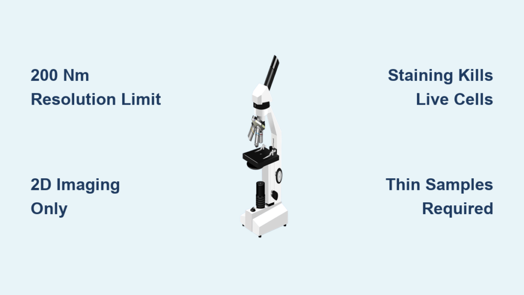
Imagine trying to examine the inner workings of a cell—mitochondria, ribosomes, or even a single virus—only to realize your microscope can’t show you the details you need. This is the daily reality for scientists, students, and medical professionals who rely on light microscopes. While these instruments are essential in biology, medicine, and education, they are fundamentally constrained by the physics of visible light. The limitations of light microscopes prevent them from resolving structures smaller than about 200 nanometers, making critical biological components invisible. Despite their affordability, ease of use, and ability to image living cells, understanding their boundaries is crucial for accurate scientific observation.
This guide breaks down every major limitation of light microscopes—from resolution caps and 2D imaging to sample prep demands and optical flaws. You’ll learn not only what these limits are but why they exist, how they affect real-world observations, and when it’s time to switch to more powerful tools like electron microscopy.
Resolution Blocked by Light Diffraction
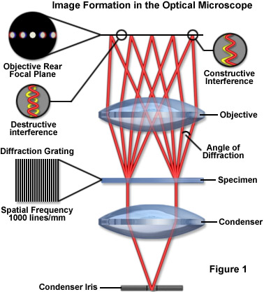
The biggest weakness of any light microscope isn’t poor lenses or weak lighting—it’s the fundamental physics of light itself. No matter how advanced the optics, the diffraction of visible light imposes a hard ceiling on what can be seen.
Abbe’s Diffraction Limit and Why It Matters
In 1873, physicist Ernst Abbe discovered that the maximum resolution of a light microscope is limited by the wavelength of light and the numerical aperture (NA) of the objective lens. Because light waves bend around tiny structures, two points closer than approximately 200 nm cannot be distinguished as separate. This is known as Abbe’s diffraction limit—a physical law, not a technical flaw.
As a result:
– Viruses (20–300 nm) appear blurred or completely undetectable.
– Ribosomes (~25 nm) and DNA strands (~2 nm) remain invisible.
– Cytoskeletal filaments like actin and microtubules are near or below the resolution threshold.
Even the best compound microscopes can’t overcome this barrier without specialized techniques.
How Resolution Is Calculated
The smallest resolvable distance $ d $ between two points is given by the formula:
$$
d = 0.61 \frac{\lambda}{NA}
$$
Where:
– $ \lambda $ = wavelength of light (e.g., 400 nm for blue light)
– $ NA $ = numerical aperture (up to 1.6 with oil immersion)
With optimal settings—blue light and oil immersion—lateral resolution reaches ~200 nm, while axial (depth) resolution is much worse at 500–700 nm. This means fine vertical layering inside cells remains blurred.
Airy Discs Blur What You’re Trying to See
When light passes through a microscopic object, it spreads into a ringed pattern called an Airy disc. If two such discs overlap, the structures they represent appear as one blurry spot. This diffraction effect limits clarity at high magnifications, no matter how powerful the lens.
Magnification Without Resolution Is Pointless
You can make an image as large as you want—but if there’s no additional detail, you’re just enlarging a blur. That’s the problem with high magnification in light microscopes.
Maximum Useful Magnification: 1500×
While some microscopes claim up to 2000× magnification, the useful limit is about 1500×. Beyond this, you experience empty magnification—the image gets bigger, but no new features appear.
Why? Because magnification depends on lens power, but detail depends on resolution. If the optics can’t resolve structures below 200 nm, blowing them up 2000 times won’t reveal more.
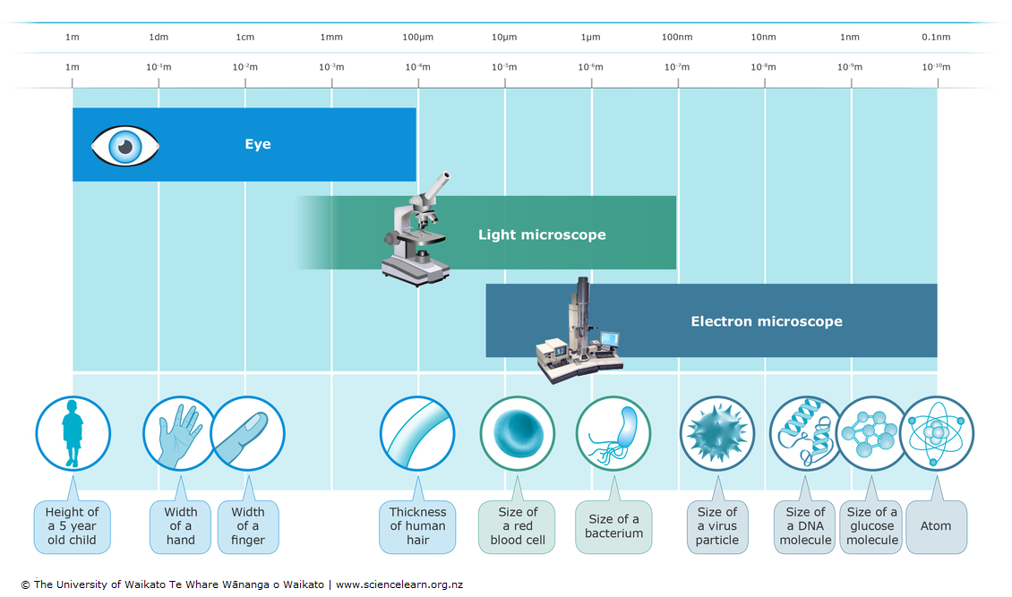
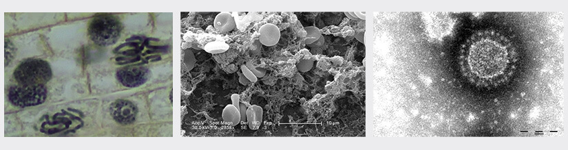
Electron Microscopes See What Light Can’t
Compare this to electron microscopes:
– Transmission Electron Microscopes (TEM) achieve <1 nm resolution and magnify up to 1,000,000×.
– Scanning Electron Microscopes (SEM) provide 3D-like surface views at 100,000× or more.
These tools are essential for studying viruses, protein complexes, and nanomaterials—areas where light microscopes simply fall short.
2D Imaging Hides 3D Structure
Standard light microscopes produce flat, two-dimensional images, making it hard to understand the true shape and depth of biological specimens.
No Depth Perception in Standard Models
Unlike our eyes or SEMs, conventional light microscopes cannot perceive depth. Structures at different levels appear stacked, with out-of-focus layers creating haze and reducing contrast.
For example:
– A neuron with complex branching may look like a tangled mess.
– Organelles in different planes blur together, obscuring spatial relationships.
Poor Axial Resolution Limits Z-Axis Clarity
While lateral resolution can reach 200 nm, z-axis (depth) resolution is only 500–700 nm. This means the microscope sees a thick “slice” vertically, blurring fine layering within cells.
Confocal Microscopy Solves the 3D Problem (With a Catch)
Confocal microscopy uses lasers and pinholes to block out-of-focus light, enabling optical sectioning and 3D reconstruction. However, this requires expensive equipment, fluorescent labeling, and complex setup—far beyond basic light microscopy.
Samples Must Be Thin and Transparent
If your specimen blocks light, a standard compound microscope won’t work. This requirement shapes how samples are prepared—and what can be studied.
Biological Tissues Need Slicing
To allow light transmission, biological samples must be cut into ultra-thin sections—5–10 micrometers thick—using a microtome. This process:
– Destroys the 3D context of tissues.
– Risks cutting through key structures.
– Is time-consuming and technically demanding.
Opaque Materials Can’t Be Viewed
Metals, ceramics, thick plastics, and dense tissues cannot be viewed with transmitted light. For these, reflected light (metallurgical) microscopes are needed—a different instrument altogether.
Staining Kills Live Cells

Many biological specimens are nearly invisible under a light microscope because they don’t absorb light. To boost contrast, scientists use stains—but this comes at a cost.
Fixation and Dyes Alter Natural State
Common stains like hematoxylin and eosin (H&E) or methylene blue require:
– Chemical fixation (e.g., formaldehyde), which kills cells.
– Permeabilization, which disrupts membranes.
As a result:
– You can’t observe dynamic processes like cell division or organelle movement.
– Structures may shrink, swell, or distort—introducing artifacts.
Live-Cell Imaging Requires Advanced Techniques
To watch living cells without staining, advanced methods are needed:
– Phase contrast: Converts subtle phase shifts into visible brightness changes.
– Differential Interference Contrast (DIC): Gives a 3D-like appearance with high contrast.
– Fluorescence microscopy: Uses glowing proteins (e.g., GFP) to label specific parts.
But these add complexity and cost—far beyond standard setups.
Surface Detail Is Poorly Resolved
Want to study the texture of a metal surface, a pollen grain, or a fractured polymer? A standard light microscope won’t give you the detail you need.
No True Topographical View
Light microscopes are designed for internal structure, not surface texture. They lack:
– Depth of field for rough surfaces.
– Resolution to see micro-relief or nanoscale roughness.
Even with reflected light, surface features appear flat and poorly defined.
SEM Wins for Surface Analysis
Scanning Electron Microscopes (SEM) excel here, offering:
– Nanoscale resolution of surface textures.
– 3D-like imaging with excellent depth perception.
– Applications in materials science, forensics, and semiconductor inspection.
For any work requiring surface topography, SEM is the gold standard.
Requires Constant Light Source
No light, no image. Unlike electron microscopes that generate their own beam, light microscopes depend entirely on external illumination.
Built-In Lamps Are Essential
Most modern scopes use:
– LEDs: Cool, long-lasting (up to 50,000 hours), energy-efficient.
– Halogen bulbs: Bright but hot, potentially damaging live specimens.
Without power, the microscope is useless—even in daylight.
Heat Can Damage Specimens
Halogen lamps emit significant heat, which can:
– Denature proteins in live cells.
– Dry out wet mounts.
– Alter physiological processes during observation.
LEDs solve this issue, but older models still pose risks.
Image Quality Degrades Without Enhancement
Even with perfect focus, raw images from light microscopes often lack contrast and clarity—especially for unstained, transparent samples.
Unstained Cells Are Hard to See
Live cells, organelles, and clear tissues scatter light weakly. Without enhancement, they appear as faint, ghostly shapes with little detail.
Contrast Techniques Add Cost
To fix this, specialized methods are used:
– Darkfield microscopy: Illuminates specimens from the side, making them glow against a dark background.
– Polarized light: Reveals birefringent materials like crystals or collagen fibers.
– Fluorescence: Tags molecules with glowing dyes for precise targeting.
But each technique requires additional optics, filters, or labels—increasing cost and complexity.
Aberrations Degrade Image Fidelity
Low-quality lenses introduce distortions:
– Chromatic aberration: Colors fringing around edges due to uneven focusing of wavelengths.
– Spherical aberration: Blurring from lens curvature imperfections.
High-end apochromatic or plan-achromatic objectives correct these issues—but at a premium price.
High Magnification Brings Practical Challenges
More power means more problems. As magnification increases, so do operational risks.
Tiny Working Distance Risks Damage
At 100× oil immersion, the space between the lens and slide is less than 0.2 mm. A slight misstep during focusing can:
– Scratch the lens.
– Crack the slide.
– Ruin the specimen.
Users must work slowly and carefully—especially with mechanical stages.
Shallow Depth of Field Requires Constant Refocusing
At high magnifications, only a thin plane is in focus. To view different layers:
– You must constantly adjust the fine focus knob.
– Thick specimens appear fragmented across multiple images.
This makes it hard to get a complete picture of 3D structures like embryos or biofilms.
Not All Specimens Are Compatible
Despite their versatility, light microscopes have strict compatibility rules.
Transmitted Light Design Limits Use
Standard compound scopes rely on light passing through the sample. This excludes:
– Metals
– Alloys
– Thick industrial materials
These require metallurgical microscopes with reflected illumination—different optics, different setup.
No Vacuum or Extreme Environment Support
Unlike electron microscopes, light scopes operate in ambient air, which allows live-cell imaging but prevents use in vacuum, high pressure, or controlled atmospheres.
When to Switch to Electron Microscopy
Light microscopes are ideal for many tasks—but not all. Knowing when to upgrade ensures accurate results.
Use Light Microscopes For:
- Observing whole cells, bacteria, and tissues
- Live-cell imaging (e.g., cell motility, mitosis)
- Clinical diagnostics (blood smears, histology)
- Educational labs and routine quality checks
Choose Electron Microscopy When:
- Studying viruses, protein complexes, or nanoparticles
- Needing ultrastructural detail (e.g., mitochondrial cristae, nuclear pores)
- Analyzing surface morphology of metals or semiconductors
- Performing 3D tomography of cellular interiors
Advanced Methods Push the Limits
Even within optical microscopy, researchers have developed ways to beat the diffraction barrier.
Super-Resolution Techniques Break the 200 nm Wall
New technologies now allow resolutions down to 30–80 nm:
– STED (Stimulated Emission Depletion): Uses a second laser to shrink the fluorescent spot.
– NSOM (Near-Field Scanning Optical Microscopy): Probes surfaces with nanoscale tips to bypass diffraction.
– Structured Illumination (SIM): Combines patterned light and computational processing to enhance resolution.
These methods are revolutionizing cell biology—but remain expensive and complex, limiting routine use.
Immersion Improves Resolution
Using oil, water, or glycerol between the objective and specimen increases the refractive index, boosting NA and cutting resolution to ~130 nm. This is standard in 100× oil immersion objectives and offers a simple upgrade path.
Summary: Key Limitations at a Glance
| Limitation | Consequence |
|---|---|
| ~200 nm resolution limit | Cannot see viruses, ribosomes, DNA |
| Empty magnification beyond 1500× | No new detail, just bigger blur |
| 2D imaging only | Poor depth perception, no 3D structure |
| Thin, transparent samples required | Thick or opaque materials excluded |
| Staining kills live cells | Dynamic processes lost; artifacts possible |
| No surface topography | Weak for materials science or texture analysis |
| Dependent on light source | Needs power; halogen heat can damage samples |
| Optical aberrations | Color fringes, blurring in low-quality lenses |
| Shallow depth of field | Frequent refocusing needed |
| Incompatible with opaque materials | Requires separate metallurgical scope |
Final Note: The limitations of light microscopes are rooted in the physics of visible light, not poor engineering. While they remain indispensable for education, diagnostics, and live-cell imaging, their resolution cap and 2D output restrict their use in modern nanoscale research. Techniques like electron microscopy and super-resolution imaging have emerged to fill these gaps. Understanding these constraints empowers scientists and students to choose the right tool—and interpret results with accuracy.


