
Microplastics under microscope are no longer just a scientific curiosity—they’re undeniable evidence of a global pollution crisis unfolding in plain sight. These synthetic fragments, smaller than 5 millimeters, have infiltrated every corner of our planet and bodies. From shimmering fibers in laundry water to glowing specks in air filters, microscopy reveals what the naked eye misses: a hidden world of persistent plastic debris. Scientists use advanced imaging to study these particles, but even basic tools can expose their presence in everyday environments. With techniques like polarized light and Nile Red staining, anyone can observe microplastics and begin to understand their scale and impact.
Seeing is believing—and understanding what you’re viewing transforms abstract concern into urgent awareness. This article guides you through proven methods for detecting microplastics, interpreting their morphology, and recognizing their sources. You’ll learn how they enter ecosystems and human tissues, the health risks they may pose, and what emerging science offers in response. Whether you’re a student, educator, or citizen scientist, observing microplastics under microscope empowers you to see the invisible and act on what you find.
See Microplastics Clearly: Best Microscopy Methods
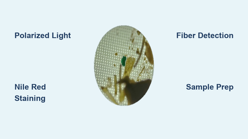
Use Polarized Light for Fiber Detection
Polarized light microscopy is one of the most effective ways to detect synthetic microfibers. By placing a polarizing filter beneath the microscope stage and another above the sample, you create a dark background that causes plastic particles to “light up” due to birefringence—a property of anisotropic materials like polyester, nylon, and acrylic.
When you rotate the polarizers, synthetic fibers display vivid rainbow hues, while natural fibers like cotton or wool remain dark. This contrast makes it easy to distinguish human-made pollutants from organic matter in complex samples such as dust, wastewater, or biological specimens. The technique is low-cost, requiring only inexpensive polarizing film, and highly accessible for educational or DIY use.
Look for uniform fiber thickness, geometric edges, and color shifts during rotation—hallmarks of synthetic origin. This method excels at identifying microfibers shed from clothing, which dominate household microplastic pollution.
Apply Nile Red Staining for Fast Screening
For rapid, reliable detection, Nile Red staining combined with fluorescence microscopy is a game-changer. The dye selectively binds to hydrophobic surfaces—like those of plastic particles—and fluoresces brightly under blue or green light (typically 450–550 nm excitation).
After filtering a sample, apply a drop of Nile Red solution, wait 1–2 minutes, then observe under fluorescence. Plastic particles glow against a dark background, making them easy to spot. This method detects particles as small as 5 µm, with ongoing improvements pushing detection toward 1 µm.
Crucially, organic debris like plant matter or hair rarely fluoresces, reducing false positives. Studies show over 90% reliability when Nile Red results are confirmed with Raman spectroscopy. While fluorescence microscopes are more specialized, this technique drastically reduces manual scanning time and increases confidence in visual identification.
Choose the Right Microscope Setup
You don’t need a high-end lab to begin. A compound light microscope at 40x total magnification (4x objective + 10x eyepiece) performs as well as stereomicroscopes commonly used in published research for initial screening.
| Magnification | Best For |
|---|---|
| 40x | Initial survey, fiber detection |
| 100x–200x | Morphology, edge analysis |
| 400x | Surface texture and fine details |
Higher magnifications reduce depth of field, making focusing difficult, especially for uneven particles. For most citizen science applications, 40x to 100x is ideal. Always use transmitted light when viewing transparent particles on filters to maximize clarity.
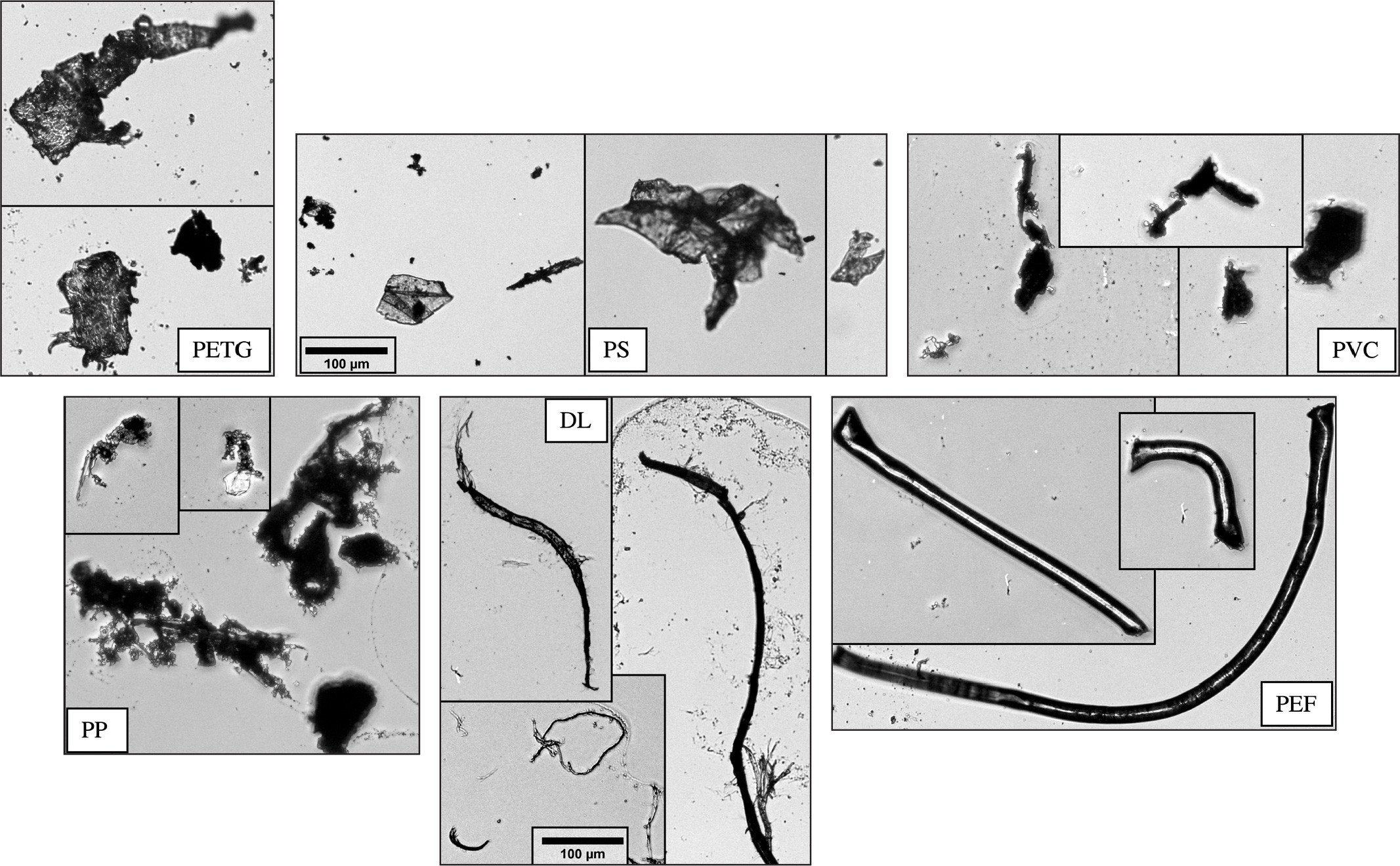
Avoid Misidentification Traps
Visual identification alone carries significant risk. Cotton fibers can mimic polyester; plant fragments may resemble plastic shards. To reduce errors, look for key synthetic indicators:
- ✅ Sharp, geometric edges
- ✅ Uniform diameter along fiber length
- ✅ Cut or melted ends (not frayed)
- ✅ No cellular structure under high power
Conversely, red flags for organic material include branching patterns, irregular thickness, and visible cell walls. Because misidentification is common, spectroscopic confirmation (FTIR or Raman) is essential for research-grade results. However, combining polarized light and staining improves accuracy even without advanced tools.
How to Collect and Prepare Samples
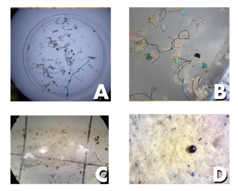
Find Microplastics in Everyday Sources
You can collect microplastics from common household sources:
– Washing machine discharge: A single load releases hundreds of thousands of microfibers.
– Navel lint: Mostly synthetic fibers shed from clothing.
– Tap and bottled water: Bottled water contains 2–3x more microplastics.
– Air filters: Capture tire wear and textile particles.
Use a coffee filter to collect fibers from laundry water. Rapid clogging indicates high microfiber load—a visible sign of pollution.
Process Samples for Clear Viewing
To isolate microplastics:
1. Digest organic matter with 30% hydrogen peroxide or KOH, heated gently (60°C) for several hours.
2. Separate minerals using sodium chloride (1.2 g/cm³) or zinc chloride (1.7 g/cm³); plastics float, sand sinks.
3. Filter the suspension through 0.45 µm cellulose nitrate membrane filters.
4. Dry and mount: Air-dry or place residue on a slide with mounting medium and cover slip.
For immediate viewing, place a drop of filtrate directly on a slide—this avoids white-on-white contrast issues.
Make Permanent Slides That Last
Preserve findings using Canada balsam or DPX mounting medium. Seal edges with nail polish and cure for 2–4 weeks in a dust-free area. Permanent slides allow long-term study and documentation.
Where Microplastics Come From and How They Spread

Tire Wear Dominates Airborne Pollution
Tire and brake wear is the largest source of airborne microplastics, especially near roads. These rubber particles become road dust and aerosolize into the air, found in both urban and rural areas. They carry heavy metals like zinc and are inhaled, depositing in lung tissue. Unlike banned microbeads, tire particles remain unregulated and a growing concern.
Clothing Sheds Synthetic Microfibers
Every wash of synthetic clothing—polyester, nylon, acrylic—releases microfibers. A single garment can shed over 1,900 fibers per wash, with fleece jackets being high-shedding. Wastewater plants capture 60–90%, but billions still enter rivers and oceans. Filters like Guppyfriend or Cora Ball reduce shedding by up to 80%.
Plastic Breakdown Creates Secondary Microplastics
Larger plastics fragment via UV radiation, mechanical abrasion, and thermal cycling, forming microplastics and nanoplastics. Even “biodegradable” plastics like PLA often fragment without fully decomposing, especially in cold marine environments.
Health Risks: What Microplastics Do in the Body
Microplastics Are in Human Tissues
They’ve been found in lungs, liver, placenta, brain, and blood. Notably, lung levels don’t increase with age, suggesting the body may clear some particles.
They Carry Toxins, Not Just Plastic
Plastics act as “toxic taxis”, adsorbing POPs (e.g., PCBs, DDT) and heavy metals. They also harbor pathogenic bacteria in biofilms. When ingested or inhaled, they can release toxins, causing inflammation and oxidative stress.
Brain and Neurological Concerns
Preliminary studies show up to 4x more microplastics in dementia patients’ brains. Researchers are investigating whether they trigger neuroinflammation or disrupt the blood-brain barrier. The European Commission is funding studies on endocrine disruption and cancer promotion.
Ecological Damage and Food Chain Entry
Marine Life Ingests Microplastics
Plankton, fish, mussels, and seabirds consume microplastics. 90% of seabirds and over 100 marine species have been found with plastic in their guts. Bioaccumulation moves plastics up the food web.
The Plastisphere: A Floating Microbial World
Plastic debris hosts the plastisphere—a unique microbial community including plastic-degrading bacteria (Ideonella sakaiensis) and pathogens (Vibrio spp.). These biofilms can spread disease.
Soil Health Is at Risk
Microplastics alter soil porosity, water retention, and microbial communities. High moisture increases leaching of toxic additives like phthalates, reducing fungal richness.
Emerging Solutions and Biotech Hope
Bacteria That Eat Plastic
- Ideonella sakaiensis: Breaks down PET using PETase.
- Engineered Vibrio natriegens: Degrades PET in saltwater at room temperature.
- PBS-degrading enzyme: Targets bioplastic polybutylene succinate.
These offer promise for bioremediation.
New Bioplastics That Actually Degrade
- LAHB + PLA blend (Kobe University): Degrades in seawater within one week.
- Carbios enzymatic recycling: Used in L’Oréal bottles, awarded Solar Impulse Pioneer Award (2021).
But most “biodegradable” plastics still come from fossil fuels, require industrial composting, and fragment into microplastics.
How to Start Your Own Microplastic Study
Gather Essential Tools
You need:
– Microscope (40x–100x)
– Polarizing film
– Slides, filters (0.45 µm), pipette
– Nile Red stain (optional)
All are affordable and accessible.
Follow a Simple Observation Protocol
- Collect sample (laundry water, navel lint)
- Filter and dry
- Mount on slide
- Use polarized light
- Stain with Nile Red (if available)
- Document shape, color, size
Compare with known fiber references.
Avoid Common Mistakes
- Don’t rely on shape alone
- Use dark backgrounds
- Label samples clearly
- Work in a clean area
Even small efforts contribute to global understanding.
Microplastics under microscope reveal a silent crisis in plain sight. They are in our water, air, food, and bodies—visible only when we choose to look. With simple tools and careful methods, anyone can detect these particles and contribute to awareness. While biotech offers hope, the most effective solution remains reducing plastic use at the source. Observe. Understand. Act. The future of our environment depends on what we do next.


