
When you switch from a 10x to a 40x objective and your specimen stays in focus with just a tiny tweak of the fine focus knob, you’re using a parfocal microscope. This essential optical feature allows users to change magnifications without losing image clarity, making microscopy faster, safer, and more efficient. In modern labs, classrooms, and imaging centers, parfocality isn’t just a convenience—it’s a standard expectation for quality microscopes.
A parfocal microscope ensures that all objective lenses share the same focal plane, so when one lens is in focus, the others remain nearly in focus when rotated into place. This eliminates the need to constantly readjust with the coarse focus—especially critical at high magnifications where even slight movements can crack a slide or damage an expensive objective. Whether you’re a student scanning a histology sample or a researcher capturing time-lapse images of live cells, this seamless transition saves time and protects equipment.
In this guide, you’ll learn how parfocality works, how to test if your microscope has it, and why it matters across scientific and imaging fields.
What Makes a Microscope Parfocal
Shared Focal Plane Across Objectives
The core of parfocality lies in precise optical alignment. Each objective lens—whether 4x, 10x, 40x, or 100x—is engineered so that its focal plane aligns exactly with those of the other objectives. This means the point at which the specimen comes into sharp focus is the same for all lenses.
When the 4x objective brings a cell into focus, switching to 10x should show that same cell still mostly clear. Only minor adjustments with the fine focus knob are needed. This consistency is not accidental—it results from strict manufacturing standards.
Parfocality is built into the design of high-quality microscopes during assembly. Manufacturers align each objective so that their optical axes converge at the same working distance above the specimen. This precision allows researchers to move from low- to high-power observation without losing their target area.
Role of Parfocal Distance
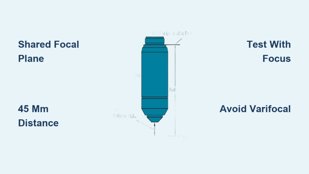
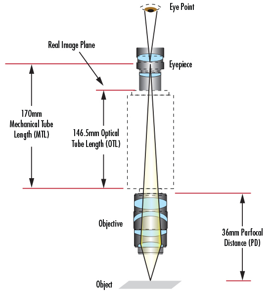
All parfocal microscopes adhere to a standard parfocal distance: the space between the bottom of the objective lens (where it screws into the nosepiece) and the specimen when in focus.
- Most modern systems, including Nikon’s UIS2 and Zeiss designs, use a 45 mm parfocal distance.
- Some specialized objectives may vary slightly, so always check manufacturer specs.
- This standard allows interchangeable objectives from the same brand to maintain parfocality.
If objectives don’t share the same parfocal distance, the image shifts out of focus when you rotate the nosepiece—defeating the purpose of a smooth magnification change. For example, inserting a 40x objective with a shorter parfocal length will cause the lens to sit too close to the slide, resulting in blurriness or even collision during use.
How to Test for Parfocality
Step-by-Step Focus Check
You can verify parfocality in under two minutes:
- Mount a prepared slide on the stage.
- Rotate to the lowest power objective (usually 4x).
- Use coarse and fine focus to sharpen the image.
- Without touching focus, rotate to the next objective (10x).
- Observe: Is the image still mostly in focus?
- Repeat up to 40x or 100x.
✅ Parfocal result: Image remains visibly clear; only fine focus tweaks needed.
❌ Non-parfocal result: Image blurs significantly; coarse adjustment required.
Most educational and research-grade compound microscopes pass this test. Budget or older models often fail, requiring full refocusing at each power.
What to Look For
- Minimal drift: A slight softening is normal, but total loss of focus is not.
- No stage crashes: If you must lower the stage drastically after switching lenses, the scope isn’t parfocal.
- Consistency across objectives: All lenses—not just adjacent ones—should behave similarly.
This test is crucial when buying used equipment or comparing models. It also helps identify misaligned objectives after handling or transport.
Why Parfocality Matters in Practice
Faster Workflow in Labs
Time adds up when refocusing at every magnification. In a clinical lab processing dozens of slides daily, parfocality can save hours per week.
- Pathologists scan tissues at low power, then zoom in on suspicious areas—without losing their spot.
- Students stay engaged instead of fumbling with focus knobs.
- Researchers tracking cell movement switch objectives mid-experiment with confidence.
Parfocality turns microscopy from a mechanical chore into a fluid observational process. Instead of constantly adjusting focus, users can maintain visual continuity, improving accuracy and reducing observer fatigue.
Reduced Risk of Slide Damage
High-magnification objectives have extremely short working distances—sometimes less than 0.1 mm. Using the coarse focus knob at 40x or 100x risks:
- Crushing the coverslip
- Scratching the lens
- Ruining rare or irreplaceable samples
A parfocal system keeps the image close to focus, so you never need the coarse knob after initial setup. This safety feature is non-negotiable in professional settings.
For instance, oil-immersion 100x objectives are particularly vulnerable. A single misstep with the coarse focus can lead to costly repairs. Parfocality minimizes that risk by ensuring the lens never plummets into the slide during magnification changes.
Essential for Digital Imaging
In digital microscopy, parfocality ensures what you see is what you capture.
- If the camera and eyepieces are not parfocal, the image may look sharp through the lens but blurry in photos.
- Many C-mount adapters include a focus adjustment ring to align the camera sensor with the eyepiece focal plane.
- Tip: Set eyepiece diopters to zero before calibrating camera focus.
For photomicrography, time-lapse videos, or publication-quality images, parfocal alignment between visual and digital outputs is mandatory. Without it, researchers risk publishing out-of-focus data or wasting time correcting focus mismatches.
Parfocality in Different Microscope Types
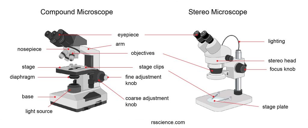
Compound Microscopes: Standard Parfocality
Most brightfield compound microscopes are parfocal by design. Brands like Olympus, Leica, and Amscope build their research and teaching models with:
- Standardized 45 mm parfocal distance
- Pre-aligned objectives
- Interchangeable lenses that maintain focus
This makes them ideal for classroom use, histology, microbiology, and clinical diagnostics. Because students often switch magnifications while learning, parfocality reduces frustration and supports faster learning curves.
Stereomicroscopes: Zoom Parfocality
In stereomicroscopes, parfocality applies across the zoom range, not discrete objectives.
- Once focused at maximum magnification, zooming out should keep the sample in focus.
- True parfocal zoom systems (e.g., Leica MZ series) require no refocusing during zoom.
- Non-parfocal stereo scopes force constant adjustments—slowing down dissection or inspection tasks.
For microsurgery, PCB inspection, or entomology, a parfocal zoom stereo microscope is indispensable. Without it, technicians lose valuable time constantly refocusing while trying to maintain hand-eye coordination.
Upgrading Non-Parfocal Systems
Adding Shims for Alignment
Some non-parfocal microscopes can be converted using thin metal or plastic shims placed between the objective and nosepiece.
- Shims adjust the objective’s position, correcting its focal plane.
- Requires trial and error to find the right thickness.
- Best done by a technician—improper shimming can cause misalignment or wobble.
However, the labor and precision involved often make this more expensive than buying a parfocal model.
When to Upgrade
Consider upgrading if:
– You frequently switch magnifications
– You use digital imaging
– You work with fragile or valuable samples
For occasional or beginner use, a non-parfocal scope may suffice. But for serious work, invest in parfocal design from the start.
Optional Parfocal: Buyer Beware
Cost-Saving Trade-Offs
Some manufacturers offer “optional parfocal” configurations:
- Base model ships with non-parfocal objectives.
- Parfocal upgrade costs extra.
- Marketed as a budget option for schools or hobbyists.
While tempting, skipping parfocality can lead to frustration and inefficiency. Always confirm whether parfocality is standard or optional before purchasing.
How to Verify
- Check product specs for “parfocal distance” or “parfocal objectives.”
- Look for phrases like “maintains focus across magnifications.”
- Contact the supplier: “Are the objectives parfocal out of the box?”
Don’t assume it’s included—many entry-level scopes cut this feature to reduce cost.
Parfocality Beyond Microscopy
Parfocal Lenses in Cinematography
In film and video, parfocal zoom lenses are prized for maintaining focus while zooming.
- Camera operators pre-focus at maximum zoom (where depth of field is shallowest), then zoom out smoothly.
- Eliminates focus breathing during dynamic shots.
- Used in documentaries, wildlife filming, and cinematic dolly zooms.
A varifocal lens would blur during zoom, requiring constant refocusing—unacceptable in professional production.
Photography and Wildlife Use
Wildlife photographers benefit from parfocal telephoto lenses:
- Focus at 600mm on a distant bird.
- Zoom to 400mm for a wider context—without losing focus.
- Critical when subjects move unpredictably.
Though rare in consumer lenses, high-end models from Canon, Nikon, and Sony offer near-parfocal performance.
Telescopes and Astronomy
Astronomers use parfocal eyepieces to switch magnifications quickly.
- Focus on a planet with a 10mm eyepiece.
- Swap to a 25mm for wider view—image stays sharp.
- Saves time during fleeting events like eclipses or meteor showers.
Parfocality here enhances observation efficiency and reduces missed opportunities.
Parfocal vs Varifocal: The Key Difference
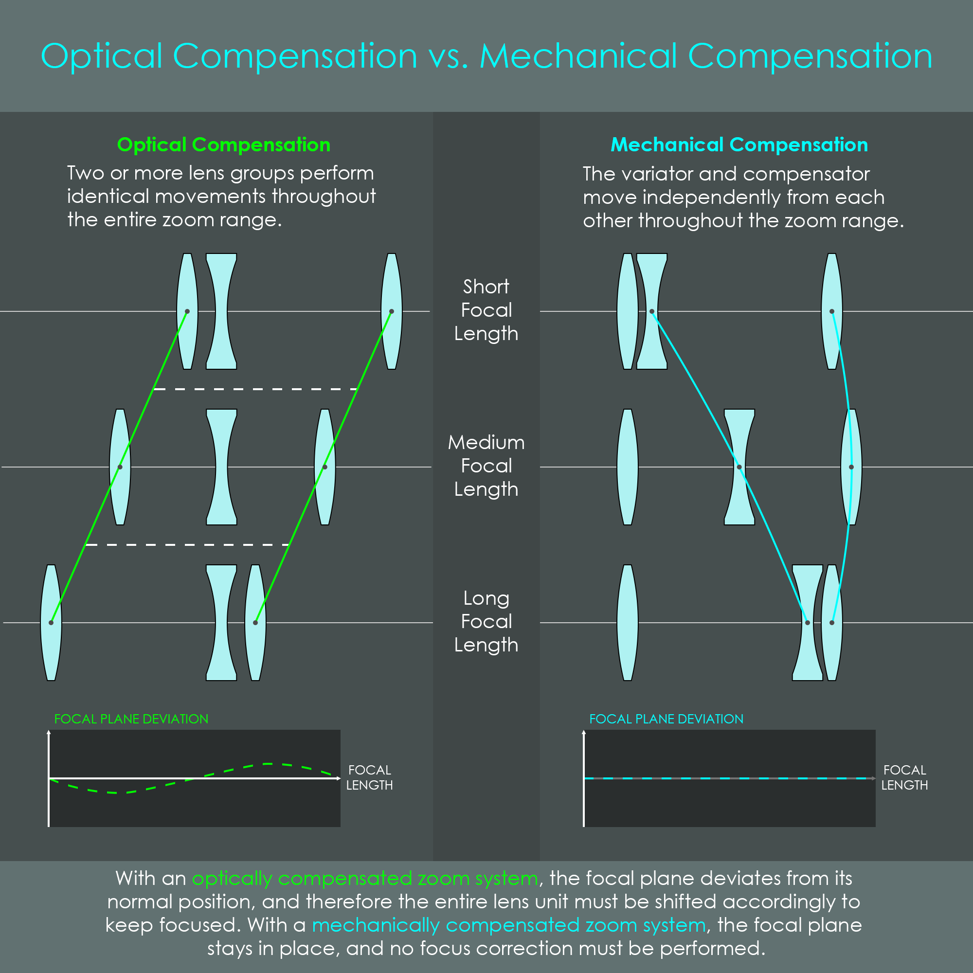
Understanding Varifocal Systems
The opposite of parfocal is varifocal—a system where changing magnification shifts the focal plane.
- Common in consumer zoom lenses with autofocus.
- Requires refocusing after every zoom or objective change.
- Rare in microscopy but found in some specialized imaging devices.
While varifocal systems offer flexibility, they sacrifice speed and precision.
Why Microscopy Avoids Varifocal Design
- Manual refocusing breaks observational flow.
- Autofocus isn’t reliable at microscopic scales.
- Risk of losing rare specimens during adjustment.
Thus, parfocal remains the gold standard in optical microscopy.
Maintaining Parfocal Performance
Handle Objectives with Care
Even a parfocal microscope can lose alignment if mishandled.
- Never force a lens into the nosepiece.
- Avoid dropping or bumping the microscope.
- Clean lenses with proper tools—scratches or debris can affect focus perception.
Regular maintenance preserves factory alignment.
Check Objective Compatibility
Mixing objectives from different manufacturers or series can break parfocality.
- Not all 45 mm parfocal distances are identical.
- Optical designs vary (e.g., infinite vs finite correction).
- Color correction, field flatness, and working distance may conflict.
Stick to matched objective sets from the same brand and series for best results.
Calibrate After Repairs
If the nosepiece or stage is serviced, realignment may be needed.
- After replacing objectives or repairing the turret, retest parfocality.
- Use a calibration slide with fine detail (e.g., stage micrometer).
- Consider professional servicing for high-end instruments.
A quick test can prevent months of inefficient use.
Applications Where Parfocality Is Critical
Live-Cell Imaging
Tracking moving cells across magnifications demands uninterrupted focus.
- Switch from overview (10x) to detail (40x) without losing the cell.
- Reduces light exposure, minimizing phototoxicity.
- Enables time-lapse studies with consistent focus.
Parfocality supports dynamic, long-term observations.
Confocal and Fluorescence Microscopy
In fluorescence work, every second under light increases photobleaching.
- Parfocal systems reduce focus adjustments, cutting exposure time.
- Essential for multi-channel imaging and z-stacks.
- Maintains alignment during automated scanning.
Focus stability equals data reliability.
Dissection and Micro-Manipulation
Stereomicroscopes used in surgery or micromanipulation rely on zoom parfocality.
- Surgeons zoom in on tissue layers without refocusing.
- Lab techs inject embryos or sort cells with continuous clarity.
- Prevents hand-eye coordination lag during precision tasks.
Here, parfocality isn’t just helpful—it’s a safety feature.
Final Note: A parfocal microscope is defined by its ability to maintain focus across magnifications, enabled by precise optical engineering and standardized parfocal distance. This feature enhances efficiency, protects samples, and supports advanced imaging. While some budget models offer parfocality as an upgrade, it remains a cornerstone of professional microscopy. Whether in education, research, or industry, choosing a parfocal system ensures smoother operation, better results, and long-term usability.


