
If you’ve ever peered into a drop of pond water and seen tiny creatures darting past, or examined the intricate structure of a leaf under magnification, you’ve experienced the power of microscopes. The types of microscope available today go far beyond basic classroom models, enabling scientists to explore everything from living cells to individual atoms. Whether you’re a student, researcher, or hobbyist, understanding the different microscope types helps you choose the right tool for your needs and appreciate how each reveals a unique layer of the microscopic world.
Microscopes work by enhancing resolution and magnification to make the invisible visible. But not all microscopes use light—some use electrons, physical probes, or even sound waves. Each type interacts with samples in distinct ways, offering specific advantages in clarity, depth, and application. This guide breaks down every major microscope type, explains how they work, and shows where they’re used—from diagnosing diseases to inspecting computer chips.
How Microscopes Work: Core Principles
Before diving into specific types, it’s essential to understand the fundamental principles that govern all microscopes: magnification, resolution, illumination, contrast, and aberration correction.
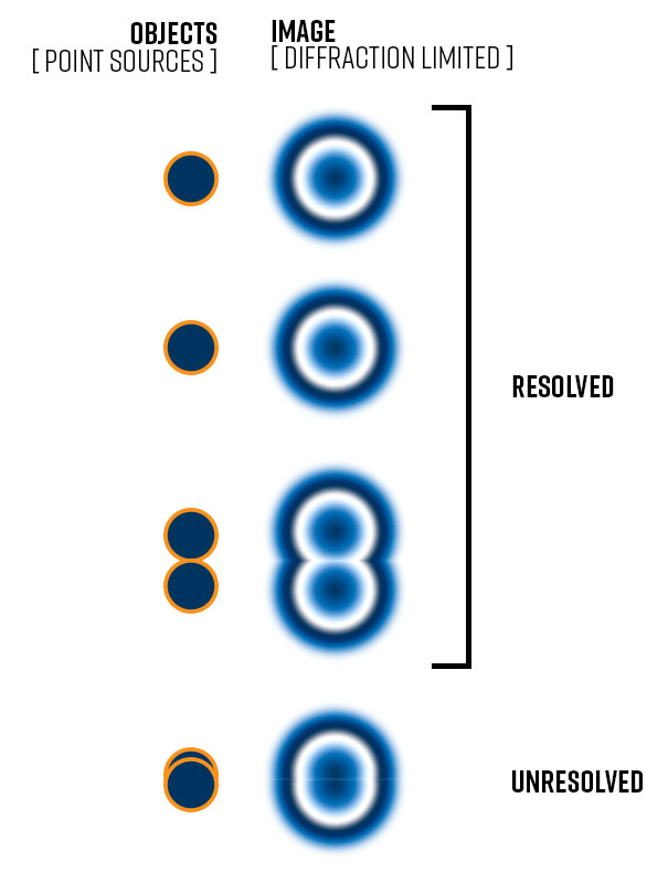
Magnification vs. Resolution
- Magnification enlarges an object’s appearance.
- Resolution determines how clearly two close points can be distinguished.
- High magnification without good resolution leads to empty magnification—a blurry image with no added detail.
For example, a compound microscope can reach 1000×, but its useful resolution maxes out around 250 nanometers due to the wavelength of visible light.
Numerical Aperture (NA) Matters
- NA measures a lens’s ability to gather light and resolve fine details.
- Higher NA = better resolution.
- Oil immersion (using oil between the lens and slide) increases NA, improving clarity at high magnifications.
Light Sources and Contrast
- Common light sources: LEDs, halogen bulbs, lasers, UV lamps.
- The iris diaphragm controls light intensity—too much washes out contrast; too little hides detail.
- Techniques like phase contrast or dark-field illumination enhance visibility of transparent specimens.
Fixing Lens Distortions
- Spherical aberration: curved images due to lens shape.
- Chromatic aberration: color fringes from light dispersion.
- Corrected using achromatic or apochromatic lenses.
Optical Microscopes: Using Light for Imaging
Optical microscopes use visible light and lenses to magnify samples. They are widely used in education, medicine, and biology because they’re accessible, easy to operate, and ideal for viewing stained or live biological specimens.
Bright-Field Compound Microscope
- Also known as a biological microscope.
- Uses transmitted light: light passes through thin, transparent samples.
- Two lens systems: objective (near sample) and eyepiece (ocular).
- Total magnification = eyepiece × objective (e.g., 10× eyepiece × 40× objective = 400×).
- Max useful magnification: 1,000× (with oil immersion).
What You Can See:
- Blood cells, bacteria, cheek cells, algae, parasites, tissue sections.
- Requires staining for contrast (e.g., methylene blue, iodine).
Where It’s Used:
- School labs, hospitals, pathology, wastewater testing, microbiology.
- Best for prepared slides—simple, fast, and effective.
Stereo Microscope for 3D Views
- Provides three-dimensional images using two separate optical paths.
- Uses reflected light, so it works on opaque or thick objects.
- Low magnification: 10× to 50×, typically 10×–40×.
- No slide preparation needed—just place the object under the lens.
Applications:
- Dissecting insects or small animals
- Inspecting circuit boards, coins, jewelry, fabrics
- Botany (flower anatomy), manufacturing quality control
Pro Tip:
Use variable zoom settings to examine large surfaces first, then focus on details.
Inverted Microscope for Live Cells
- Objective lenses are below the stage, light source above—opposite of a standard microscope.
- Designed to view cells growing in petri dishes or flasks.
- Allows long-term observation without disturbing cultures.
Key Uses:
- Live cell imaging
- In-vitro fertilization (IVF)
- Neuroscience, developmental biology, cancer research
Industrial Version:
Metallurgical inverted microscopes inspect metal surfaces, ceramics, and industrial “pucks.”
Dark-Field Microscopy: Bright Specimens on Black
- Blocks direct light with a special dark-field condenser.
- Only scattered or refracted light enters the lens.
- Results in glowing specimens against a dark background.
- Perfect for unstained, live samples.
Ideal For:
- Spiral bacteria like Treponema pallidum (cause of syphilis).
- Observing internal structures in motion within eukaryotic cells.
Limitation:
Lower resolution due to reduced light—best combined with other techniques.
Phase-Contrast: See Live Cells Without Staining
- Converts invisible phase shifts in light (caused by transparent cells) into visible contrast.
- Eliminates need for chemical stains—keeps cells alive during imaging.
- Developed by Frits Zernike, Nobel Prize winner (1953).
Applications:
- Cell division cycles
- Bacterial endospore detection
- Microbial motility studies
- Plant and animal cell morphology
Why It’s Important:
Enables real-time observation of living processes—like watching a cell divide without killing it.
DIC Microscope: 3D-Like Imaging
- Short for Differential Interference Contrast (also called Nomarski microscopy).
- Uses polarized light, prisms, and beam splitters to create high-contrast, pseudo-3D images.
- Developed in 1955 by Georges Nomarski.
Advantages:
- Sharp, shadowed appearance mimics 3D structure.
- Excellent for transparent, unstained biological samples.
Common Uses:
- Tissue culture monitoring
- Single-celled organism imaging
- Embryo development studies
Fluorescence Microscopy: Tag Specific Structures
- Uses fluorescent dyes or proteins that glow when hit by specific light (often UV or blue).
- Filters separate excitation light from emitted fluorescence.
- Enables molecular-level targeting.
Common Fluorescent Agents:
- DAPI: binds DNA (blue glow)
- Acridine orange: stains nucleic acids
- Green Fluorescent Protein (GFP): genetically encoded tag
- Immunofluorescence: antibody-linked fluorophores
Applications:
- Detecting Mycobacterium tuberculosis
- Live/dead bacterial assays
- Tracking gene expression in real time
- Neural circuit mapping
Expert Note:
Fluorescence fades over time (photobleaching)—limit exposure to preserve signals.
Confocal Laser Scanning Microscope (CLSM)
- Advanced form of fluorescence microscopy.
- Uses a laser beam and pinhole aperture to block out-of-focus light.
- Scans sample point-by-point, layer-by-layer.
- Builds optical sections for 3D reconstruction on a computer.
Benefits:
- Eliminates blur from thick specimens.
- Minimizes photodamage—ideal for live tissue.
- Generates high-resolution 2D and 3D images.
Uses:
- Cancer diagnosis (cervical, skin)
- Neurobiology (e.g., C. elegans embryos)
- Multilayered tissue imaging
Example:
A Caenorhabditis elegans embryo with somatic cells in red, gut granules in yellow—reveals internal development in stunning detail.
Polarizing Microscope: Analyze Crystals and Minerals
- Uses polarized light with a polarizer (below) and analyzer (above).
- Detects birefringent materials—those that split light into two rays.
- Reveals crystal structure, stress patterns, and molecular alignment.
Applications:
- Geology and petrology (rock/mineral ID)
- Pharmaceutical crystallography
- Urine analysis, especially amyloid deposits (show apple-green birefringence)
- Polymer and coating analysis
Example:
Vitamin C crystals imaged at 200×—reveals intricate geometric patterns.
Comparison Microscope: Side-by-Side Forensic Analysis
- Allows simultaneous viewing of two specimens through one eyepiece.
- Uses a split optical path and dual objectives.
Main Use:
- Forensics: compare bullets, cartridge casings, tool marks, fibers, documents.
- Critical in firearms identification—detecting unique striations from gun barrels.
Real-World Impact:
Helps link weapons to crimes with microscopic precision.
Digital Microscope: Image Capture and Analysis
- Replaces or adds to eyepieces with a CMOS or CCD camera.
- Displays image on a computer monitor.
- Enables measurement, annotation, storage, and sharing.
Where It’s Used:
- Education (classroom projection)
- Medical diagnostics
- Manufacturing inspection
- Forensic documentation
Bonus:
Software tools allow time-lapse imaging, particle counting, and dimensional analysis.
USB Microscope: Portable and Simple
- Low-cost digital microscope connected via USB.
- No eyepiece—image appears directly on screen.
- Magnification typically up to 40×.
Best For:
- Coin, banknote, and circuit board inspection
- Textile and fiber analysis
- Field use (e.g., flat rocks, stamps, documents)
Limitation:
Lower resolution than lab-grade models—but highly accessible.
Electron Microscopes: Seeing at the Nanoscale
Electron microscopes use electron beams instead of light, achieving far higher resolution—down to 0.1 nanometers. Electrons have much shorter wavelengths than light, allowing visualization of viruses, DNA strands, and even atoms.
But they require vacuum environments, complex preparation, and are not suitable for live imaging.
Transmission Electron Microscope (TEM)
- Electrons pass through ultra-thin samples (<100 nm thick).
- Denser areas block electrons (appear dark); lighter areas transmit (appear bright).
- Produces high-contrast 2D images of internal structures.
Sample Prep:
- Thin sectioning with ultramicrotome.
- Staining with heavy metals (e.g., osmium, lead).
History:
- First built by Ernst Ruska and Max Knoll (1931).
- Ruska won Nobel Prize (1986).
What You Can See:
- Viruses (20–300 nm), DNA (2 nm), ribosomes, chloroplast thylakoids, cell organelles.
- Nanoparticles, semiconductor layers.
Applications:
- Virology, ultrastructural biology, materials science, nanotechnology.
Example:
Bacteriophage structure revealed in full detail—head, tail, fibers.
Scanning Electron Microscope (SEM)
- Electrons scan the surface in a raster pattern.
- Detects secondary or reflected electrons to build a 3D-like image.
- Can image bulk, non-thin samples.
Sample Prep:
- Coating with gold or carbon if non-conductive.
- Must be dry and stable in vacuum.
Magnification:
- Up to ~100,000× (e.g., E. coli at 17,500×).
Applications:
- Surface topography of insects, pollen, blood cells, tardigrades
- Fracture analysis in materials
- Electronics failure inspection
- Geology, forensics
Example:
A water bear (tardigrade) imaged with astonishing texture and depth—resembles a sci-fi creature.
Scanning Probe Microscopes: Atomic-Level Touch

Instead of light or electrons, these microscopes use a physical probe to scan surfaces at atomic scales. They bypass diffraction limits and provide topographic maps based on interactions like force or electrical current.
Atomic Force Microscope (AFM)
- A sharp tip on a cantilever scans the surface.
- Measures deflection from atomic forces (van der Waals, electrostatic).
- A laser detects cantilever movement.
Advantages:
- Works in air, liquid, or vacuum.
- Can image non-conductive samples.
- Measures mechanical, magnetic, and electrical properties.
Applications:
- DNA, plasma membrane proteins, nanomaterials
- Drug delivery systems, biosensors
Example:
Visualizing individual membrane proteins—reveals how they cluster and function.
Scanning Tunneling Microscope (STM)
- Uses quantum tunneling: electrons jump between a sharp metal tip and a conductive surface.
- Tunneling current changes with distance—used to map surface topography.
- Requires conductive samples and ultra-high vacuum.
History:
- Invented by Gerd Binnig and Heinrich Rohrer (1981–1983) at IBM.
- Shared Nobel Prize (1986).
What It Can Do:
- Image atomic arrangements on surfaces.
- Manipulate individual atoms (used in nanotechnology).
Example:
DNA double helix visualized at atomic scale—confirms molecular models.
Near-Field Scanning Optical Microscope (NSOM/SNOM)
- Combines AFM mechanics with optical microscopy.
- A nanoscale aperture in a fiber tip delivers light very close to the sample.
- Captures localized optical properties beyond diffraction limit.
Use Case:
- Sub-cellular imaging with optical specificity at nanoscale.
Specialized and Emerging Microscopes
Beyond traditional categories, advanced technologies push imaging further—into quantum realms, 3D tomography, and non-destructive testing.
Acoustic Microscope: See Inside Without Cutting
- Uses high-frequency ultrasound (like SONAR).
- Sound waves reflect off internal boundaries.
- Differences in acoustic impedance create contrast.
Key Feature:
- Non-destructive—no need to open or damage components.
Applications:
- Detecting cracks, voids, delamination in ICs, composites, aerospace parts
- Failure analysis in electronics
Industry Use:
Essential in semiconductor packaging and quality assurance.
X-ray Microscope: 3D Imaging Without Staining
- Uses soft X-rays to penetrate biological and material samples.
- Can perform micro-CT scans for 3D reconstructions.
- Does not require chemical fixation—preserves natural state.
Applications:
- Biological tomography
- Bone and tooth structure analysis
- Material porosity studies
Future:
Research into hard X-ray optics aims to image deeper structures.
Super-Resolution Microscopes: Beat the Diffraction Limit
- Overcome the 250 nm resolution barrier of light microscopes.
- Techniques include:
- STED (Stimulated Emission Depletion): narrows fluorescence spot for sharper images.
- SIM (Structured Illumination): doubles resolution using patterned light.
- Enables near-electron-microscope clarity with fluorescence.
Nobel Recognition:
Stefan Hell, Eric Betzig, William Moerner won the 2014 Chemistry Nobel for single-molecule imaging.
Impact:
Now used in neuroscience, immunology, cancer research.
Quantum Microscope: Image with Minimal Damage
- Uses entangled photons—one infrared photon illuminates sample, its visible twin is detected.
- Reduces light exposure—ideal for light-sensitive biological samples.
Developed:
By Australian engineers (2013).
Principle:
- Photon-sparse imaging—gathers data without damaging delicate structures.
- Enables long-term observation of photosensitive cells.
Choosing the Right Microscope
| Need | Recommended Microscope |
|---|---|
| Basic biology lab | Compound (bright-field) |
| Live cell imaging | Phase-contrast, Inverted, Confocal |
| Dissection or QC | Stereo microscope |
| Molecular labeling | Fluorescence, Confocal |
| Virus or organelle | TEM |
| Surface texture | SEM |
| Atomic-scale detail | STM, AFM |
| Crystal analysis | Polarizing microscope |
| Electronics inspection | Acoustic, SEM |
| Field or classroom | USB, Digital microscope |
Final Thoughts: The Future of Microscopy
The types of microscope available today reflect centuries of innovation—from van Leeuwenhoek’s single-lens marvels to quantum-enabled imaging. While optical microscopes remain the workhorses of education and medicine, electron and scanning probe microscopes unlock the nanoworld. Meanwhile, super-resolution, acoustic, and quantum microscopes are redefining what’s possible.
No single microscope does it all. Each has strengths shaped by physics and purpose. By matching the tool to your sample and goal, you gain not just magnification—but insight.
Whether you’re diagnosing disease, designing new materials, or simply curious about life’s hidden layers, there’s a microscope built to reveal it.


