
Peering into a microscope and spotting a tightly packed sheet of cells with no blood vessels in sight? You’re almost certainly looking at epithelial tissue—one of the four primary tissue types in the human body. This highly organized, avascular layer forms the frontline defense and functional interface in nearly every organ system. Whether it’s shielding your skin from UV damage, absorbing nutrients in the small intestine, or filtering gases in the lungs, epithelial tissue reveals a striking harmony between structure and function under microscopic examination.
Identifying epithelial tissue accurately requires more than just recognizing cell clusters—it demands attention to specific histological clues: cell shape, number of layers, nuclear positioning, and surface specializations like cilia or microvilli. These features allow students, histologists, and medical professionals to classify epithelia precisely and infer their physiological roles. In this guide, we’ll walk you through how to confidently identify epithelial tissue under the microscope, avoid common misidentifications, and understand what each structural detail tells you about its function. From the trachea to the bladder, you’ll learn how to decode real-world examples using key morphological patterns.
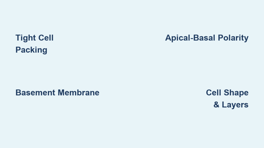
Identify the Defining Microscopic Traits of Epithelial Tissue
Before classifying any epithelium, confirm you’re actually looking at epithelial tissue by checking for its hallmark characteristics visible under light microscopy.
Look for Dense Cell Packing with Minimal Gaps
Epithelial cells are tightly packed, forming continuous sheets with little to no extracellular matrix between them. This creates a seamless barrier ideal for protection, absorption, or secretion. Unlike connective tissue—which appears fibrous and scattered—epithelium looks like a wall of cells edge-to-edge. When examining a slide, focus on:
– Cells touching laterally with sharp, defined borders
– Minimal visible space between adjacent cells
– Uniform arrangement, especially in cuboidal or columnar types
This compact organization supports selective permeability and mechanical strength depending on location.
Locate the Basement Membrane
All epithelial tissues rest on a basement membrane, a thin, pink-staining line (in H&E-stained slides) that separates the epithelium from underlying connective tissue. Though not a cellular layer, it’s rich in collagen and laminin and serves as a critical anchor. Under the microscope:
– It appears as a smooth, dark line beneath the deepest cell layer
– Helps determine epithelial orientation when the tissue isn’t at the slide’s edge
– Is essential for nutrient diffusion since epithelia lack blood vessels
Never mistake the basement membrane for connective tissue—its presence confirms you’re viewing true epithelium.
Assess Apical-Basal Polarity
Epithelial cells exhibit polarity: distinct apical (top) and basal (bottom) surfaces.
– The apical surface faces the lumen or external environment (e.g., gut cavity or airway)
– The basal surface attaches to the basement membrane
– Nuclei are typically aligned toward the basal side
This polarity is most obvious in columnar epithelia, where elongated nuclei sit near the base while the cell body extends upward.
Classify Epithelium by Layers and Cell Shape

The standard classification system combines number of layers and superficial cell shape to name epithelial types precisely.
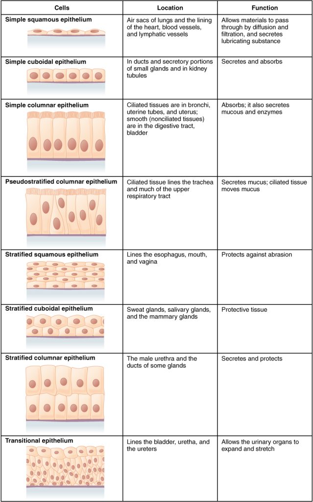
Simple Epithelium: One Layer for Efficient Exchange
Simple epithelium consists of a single layer of cells, all resting on the basement membrane. It’s found where rapid diffusion, absorption, or secretion occurs.
Simple Squamous Epithelium
Flattened cells with thin cytoplasm and central, oval nuclei. Nuclei resemble scattered pancakes or “fried eggs” (yolk = nucleus, white = cytoplasm). Found in alveoli, kidney glomeruli, and blood vessels, it minimizes diffusion distance for efficient gas and fluid exchange.
Simple Cuboidal Epithelium
Cube-shaped cells with round, centrally located nuclei. Appears as a uniform, tile-like pattern. Present in kidney tubules and glandular ducts, it supports secretion and absorption.
Simple Columnar Epithelium
Tall, rectangular cells with basal nuclei. Often contains goblet cells (clear, mucus-secreting cells) and microvilli (fuzzy brush border in the intestine). Found in the stomach, intestines, and gallbladder, it specializes in nutrient uptake and mucus production.
Pseudostratified Columnar Epithelium: One Layer That Looks Layered
Despite appearing multilayered, all cells touch the basement membrane, making it a type of simple epithelium. Characterized by nuclei at multiple levels, cilia, and goblet cells, it lines the trachea and bronchi. Its mucociliary escalator traps and removes debris from the airways.
Stratified Epithelium: Multiple Layers for Durability
Stratified epithelia have two or more layers, with only basal cells attached to the basement membrane. Named by the shape of superficial cells.
Stratified Squamous Epithelium
Basal layers contain dividing cuboidal cells; surface cells are flat. Two subtypes:
– Non-keratinized: Surface cells have nuclei; found in the mouth and esophagus
– Keratinized: Outer layers are dead, filled with pink-staining keratin; no nuclei; found in the skin epidermis
Provides strong protection against abrasion and dehydration.
Transitional Epithelium (Urothelium)
Unique to the urinary system, it changes shape with organ distension. In a relaxed bladder, surface cells are dome-shaped (“umbrella cells”), sometimes binucleated. When stretched, they flatten. This elasticity allows the bladder to expand without breaking its barrier.
Determine Orientation in Histological Slides
Don’t assume epithelium is always on top. Orientation depends on how the tissue was sectioned.
Find the Epithelial Side Using Morphological Clues
Instead of relying on position, use structural cues:
– Look for tight cell sheets adjacent to a basement membrane
– Identify polarity: nuclei clustered basally, free apical surface
– Check for specialized features: cilia, microvilli, or keratin
Examples:
– Trachea: Epithelium lines the inner lumen (center of slide)
– Skin: Epidermis on top, dermis below
– Bladder: Epithelium on inner surface, even if rotated
Use Organ Context to Narrow Identification
Knowing the tissue source helps identify the epithelium. Key examples:
| Organ | Epithelial Type | Key Clue |
|---|---|---|
| Small intestine | Simple columnar with brush border | Goblet cells + microvilli |
| Esophagus | Non-keratinized stratified squamous | Multiple layers, no keratin |
| Skin | Keratinized stratified squamous | Pink keratin layer, no nuclei on surface |
| Trachea | Pseudostratified ciliated columnar | Cilia + goblet cells + staggered nuclei |
| Urinary bladder | Transitional epithelium | Umbrella cells, binucleated surface cells |
| Kidney tubule | Simple cuboidal | Uniform cubes, central nuclei |
Optimize Microscopy Technique for Clear Viewing
Proper setup ensures accurate identification.
Set Up the Microscope Correctly
- Start low: Use 4x or 10x to locate the tissue
- Center specimen: Move slide so epithelium is over light path
- Focus safely: Lower objective while watching from the side, then raise stage slowly using coarse knob
- Use fine focus only at higher magnifications
- Increase gradually: Rotate to 20x, then 40x
- Avoid 100x oil immersion unless necessary—cleaning is cumbersome
Never use coarse focus at high power—it can crack the slide.
Choose the Right Magnification
- 100x total (10x objective): Overview of tissue structure
- 200x total (20x objective): Best for identifying layers and cell shape
- 400x total (40x objective): Ideal for nuclei, cilia, microvilli, keratin
For most identifications, 200x–400x provides optimal detail.
Avoid Common Misidentification Mistakes

Even experienced learners make errors.
Don’t Confuse Pseudostratified with Stratified
- Pseudostratified: All cells reach basement membrane; nuclei at different heights
- Stratified: Only basal cells touch base; superficial cells may lack nuclei
Clue: If every cell extends to the base, it’s pseudostratified.
Distinguish Keratinized vs. Non-Keratinized
- Keratinized: No nuclei in outer layer, bright pink staining (skin)
- Non-keratinized: Surface cells have nuclei (esophagus)
Location is key: skin = keratinized; internal moist surfaces = non-keratinized.
Watch for Artifacts
- Folds or tears can mimic multiple layers
- Poor staining may obscure nuclei
- Oblique sectioning distorts cell shape
Always examine multiple fields of view.
Correlate Structure with Function
The microscope reveals physiology in action.
- Simple squamous (alveoli): Thin barrier enables rapid gas exchange
- Simple columnar (intestine): Tall cells house organelles for absorption and secretion
- Stratified squamous (skin): Layers resist friction and pathogens
- Transitional (bladder): Stretchable lining accommodates urine volume changes
Each structural detail reflects a functional adaptation.
Practice with Unlabeled Images
Sharpen skills using histology atlases with unlabeled photomicrographs of trachea, esophagus, colon, and bladder. Challenge yourself to:
– Identify epithelial type
– Determine tissue source
– Explain function
– Note magnification
Then verify answers to reinforce learning. Build a personal study guide with labeled drawings and notes to master pattern recognition.
Final Note: Mastering epithelial tissue under the microscope hinges on linking structure to function. By focusing on cell shape, layering, polarity, and specialized features—and using proper technique—you can confidently identify any epithelium. With practice, these patterns become intuitive, laying the foundation for success in histology, medicine, and biomedical sciences.


