
When you peer through a microscope, the image you see—sharp, detailed, and magnified—is primarily shaped by one crucial component: the objective lens. This small but powerful optical system sits closest to the specimen and is responsible for capturing light and forming the first, real image in the imaging chain. Without a high-quality objective lens, even the most advanced microscope fails to deliver clear results. Understanding the objective lens microscope definition, its role, and how it varies across types is essential for anyone working in biology, medicine, materials science, or industrial inspection.
The objective lens doesn’t just magnify—it determines resolution, contrast, brightness, and depth of field. Its performance is governed by key specifications like numerical aperture (NA), magnification, working distance, and aberration correction. Whether you’re using a basic school microscope or a high-end fluorescence imaging system, the objective lens sets the upper limit on what you can see. In this guide, we’ll break down everything you need to know about objective lenses—from core functions and technical specs to types, selection criteria, and care tips—so you can make informed decisions and get the most out of your microscopy work.
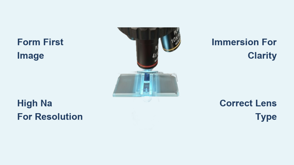
First Image Formation: The Heart of Microscopy
Why the Objective Lens Matters Most
The objective lens performs the critical task of forming the first real image of the specimen. Unlike the eyepiece, which only magnifies an already-formed image, the objective gathers light directly from the sample and focuses it into a sharp, inverted image at the intermediate image plane. This image is then either viewed through the ocular lens or captured by a digital camera.
Because all optical information originates here, any loss in detail—such as blurring, chromatic distortion, or low contrast—cannot be recovered later in the imaging chain. That’s why the objective lens is often referred to as the “heart” of the microscope. If the initial image lacks resolution or fidelity, no amount of digital enhancement can restore what was never captured.
This foundational role makes the objective lens the single most important factor in determining image quality. Even with a top-tier camera or eyepiece, a poorly performing objective will limit your ability to resolve fine structures, whether you’re studying cellular organelles or microchip circuits.
Light Collection and Resolution: The Role of Numerical Aperture
How NA Defines Image Quality
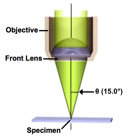
Two of the objective lens’s most vital functions are collecting light efficiently and resolving fine details. The numerical aperture (NA) is the key metric that governs both. NA measures the lens’s ability to gather light and resolve small specimen details at a fixed distance.
It’s calculated using the formula:
NA = n × sin(θ)
Where:
– n = refractive index of the medium (air, oil, water)
– θ = half-angle of the maximum light cone entering the lens
Higher NA values mean better resolution and brighter images. For example, a 100x oil-immersion objective with NA 1.4 can resolve structures as small as 240 nanometers, far beyond what a dry lens with NA 0.95 can achieve.
Resolution (R) is determined by:
R = 0.61 λ / NA
With λ being the wavelength of light (~550 nm for visible light). This means resolution improves as NA increases—making NA more important than magnification alone.
However, higher NA also brings trade-offs: reduced depth of field and smaller field of view, requiring precise focusing and stable sample positioning.
Magnification Levels: What the Numbers Mean
Scanning, Low Power, High Power, and Oil Immersion
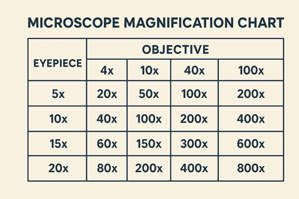
Magnification indicates how much larger the specimen appears. It’s marked on the lens barrel (e.g., 4x, 10x, 40x, 100x). Total magnification is calculated by multiplying the objective value with the eyepiece (e.g., 10x eyepiece × 40x objective = 400x total).
Common magnification categories include:
– Scanning (2x–4x): Used for initial sample location; wide field of view
– Low power (10x): General observation; ideal for large structures
– High power (40x): Detailed cellular imaging
– Oil immersion (100x): Subcellular resolution; requires immersion oil
Crucially, higher magnification does not guarantee better detail—resolution depends on NA. A 60x oil lens with NA 1.4 may outperform a 100x dry lens with NA 0.95 in clarity and fine detail.
Working Distance and Field of View: Practical Imaging Considerations
How Close Can You Get?
Working distance (WD) is the space between the front lens and the specimen when in focus. It decreases as magnification and NA increase:
– 4x objective: Up to 41 mm WD
– 100x oil objective: Less than 0.2 mm WD
Long working distance (LWD) and ultra-long working distance (ULWD) objectives are essential for:
– Live-cell imaging
– Micromanipulation (e.g., patch clamping)
– Industrial inspections where tools must access the sample
How Much Can You See?
The field of view (FOV) is the diameter of the visible area, calculated as:
FOV = Eyepiece Field Number / Objective Magnification
For example, a 22 mm field number eyepiece with a 40x objective gives a FOV of 0.55 mm. As magnification increases, the FOV shrinks—so always start with a low-power objective to locate your sample.
Objective Lens Types: From Achromat to Apochromat
Color Correction Levels
Chromatic aberration causes different colors to focus at different planes. Objective lenses correct this to varying degrees:
| Type | Color Correction | Best For |
|---|---|---|
| Achromat | Red and blue corrected; violet out of focus | Routine brightfield |
| Semiapochromat (Fluorite) | Better correction; uses fluorite glass | Fluorescence, multicolor imaging |
| Apochromat | Full correction across three wavelengths | High-resolution research |
Apochromats offer superior color fidelity but are more complex and expensive, often containing over 15 lens elements.
Plan Objectives: Flat Field Imaging
Standard objectives suffer from field curvature—only the center is sharp. Plan objectives correct this, delivering a flat image across the entire field.
- Plan Achromat: Affordable flat-field option
- Plan Fluor: Balanced for fluorescence
- Plan Apochromat: Top-tier clarity and flatness
Plan lenses are essential for digital imaging, photomicrography, and measurement applications.
Immersion Objectives: Pushing Resolution Limits
Oil, Water, and Glycerol Immersion Explained
Immersion objectives use a liquid between the lens and specimen to reduce light refraction and increase NA.
- Oil-immersion (NA up to 1.45): Used with 100x objectives; requires immersion oil (n ≈ 1.51)
- Water-immersion (NA up to 1.2): Ideal for live-cell imaging
- Glycerol-immersion (NA ~1.3): Matches mounting media in deep-tissue imaging
Using oil incorrectly—or not at all—leads to severe image degradation due to light scattering at air-glass interfaces.
Optical Design: Finite vs Infinity-Corrected Systems
Which System Does Your Microscope Use?
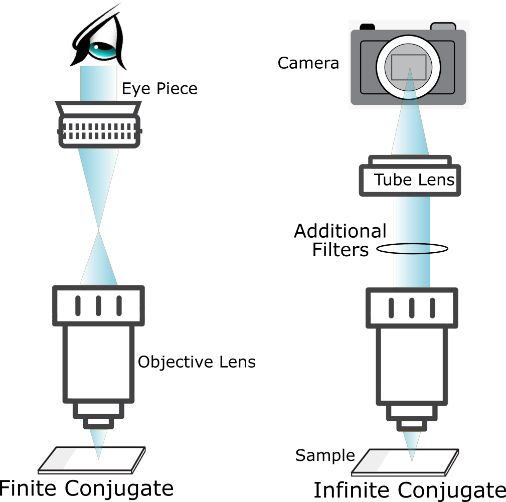
Microscopes use two main optical designs:
| Type | Features | Applications |
|---|---|---|
| Finite Conjugate | Fixed tube length (160 mm); light converges directly | Older models; basic systems |
| Infinity-Corrected | Emits parallel rays; requires tube lens | Modern research microscopes |
Infinity-corrected systems allow insertion of filters, prisms, and beam splitters, making them ideal for fluorescence, DIC, and confocal microscopy.
Specialized Objectives for Advanced Applications
Matching the Lens to the Technique
Different microscopy methods require dedicated objectives:
- Phase Contrast: Internal phase ring enhances contrast in transparent, unstained cells
- DIC (Nomarski): Works with prisms to create 3D-like contrast in live samples
- Fluorescence: High transmission in UV-Vis range; low autofluorescence
- TIRF: Very high NA (≥1.45); excites fluorophores near the cell membrane
- Polarization: Strain-free design prevents birefringence artifacts
- Industrial (Metallurgical): Designed for uncovered, reflective surfaces like metals
Each type ensures optimal performance for its specific imaging modality.
How to Read Objective Lens Markings
Decoding the Engraved Specs
Every objective has labeled specifications. Example:
Plan Achromat 10x/0.25 ∞/0–2 mm 160/DIN
- Plan Achromat: Flat field with chromatic correction
- 10x: Magnification
- 0.25: Numerical aperture
- ∞: Infinity-corrected
- 0–2 mm: Cover slip correction range
- 160/DIN: Finite tube length standard
Other common labels:
– Oil, Water, Glyc: Required immersion medium
– APO: Apochromatic correction
– LWD/ULWD: Long working distance
– UV/NIR: Optimized for specific wavelengths
Always verify compatibility with your microscope and application.
How to Choose the Right Objective Lens
Match the Lens to Your Sample
Ask these questions:
– Live cells? → Water immersion, long working distance
– Fluorescent labels? → High-NA, low-autofluorescence lens
– Unstained tissue? → Phase contrast or DIC
– Metals or circuits? → Industrial objective, no cover slip
Prioritize NA over magnification for resolution. A 60x oil objective with NA 1.4 often outperforms a 100x dry lens.
Ensure compatibility with:
– Infinity vs finite systems
– Thread standard (RMS 20.32 mm common)
– Parfocal distance (for smooth lens switching)
Care and Maintenance: Protect Your Investment
Cleaning Without Damage
- Dust: Use a clean air blower—never breathe on the lens
- Smudges: Use lens paper and 70% ethanol
- Immersion oil: Wipe immediately; use isopropyl alcohol for dried residue
Never use:
– Paper towels
– Acetone (damages coatings)
– Fingers (oils degrade performance)
Handling Tips
- Always handle by the barrel
- Store with the lowest-power objective in place
- Use dust covers when idle
- Avoid bumping the front lens—especially high-NA oil objectives
A scratched or dirty front lens permanently degrades image quality.
The Future of Objective Lenses
AI and Smart Optics
Modern systems integrate machine learning to:
– Enhance resolution beyond optical limits
– Reduce noise in low-light imaging
– Automate focus and region detection
Emerging smart objectives may include:
– Embedded sensors for real-time feedback
– Auto-identification via RFID
– Adaptive optics that correct sample-induced aberrations
These innovations aim to simplify complex workflows and improve reproducibility.
Final Note: The objective lens is far more than just a magnifier—it’s the defining element of a microscope’s performance. From its role in image formation to the nuances of NA, correction types, and immersion media, every specification impacts what you can see and how clearly. By understanding the objective lens microscope definition and its practical implications, you can select the right lens for your needs, maintain it properly, and unlock the full potential of your microscope—whether you’re studying cells, materials, or microcircuits.


