
Peering at a neuron under microscope reveals one of nature’s most intricate creations—the fundamental unit of thought, sensation, and movement. Unlike typical cells, neurons possess a striking, tree-like structure that reflects their specialized role in transmitting information across the nervous system. With magnification, what was once invisible becomes a detailed landscape of branching dendrites, a central cell body, and a long, cable-like axon. These structures aren’t just visually complex—they are precisely engineered for rapid communication, learning, and adaptation. Whether using light microscopy to identify stained cell bodies or electron microscopy to explore synaptic junctions, each imaging technique uncovers a new layer of neural architecture. This guide explores exactly what you observe when examining a neuron under the microscope, how scientists enhance these views, and how microscopic structure directly shapes brain function.
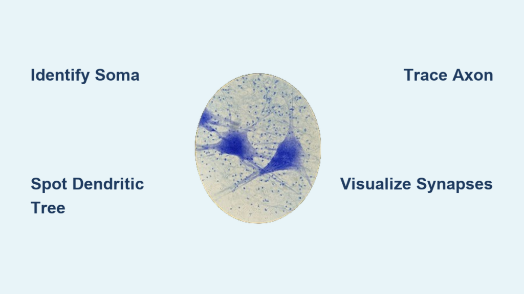
Identify the Core Components of a Neuron
Locate the Cell Body (Soma)
The soma, or neuronal cell body, is usually the first feature visible under the microscope. It appears as a large, round or oval structure with a prominent, dark-staining nucleus at its center. Inside the nucleus, a smaller, intensely stained spot—the nucleolus—may be visible, signaling active ribosome production. The soma houses essential organelles like mitochondria, Golgi apparatus, and rough endoplasmic reticulum (RER), all vital for protein synthesis and energy generation.
When tissue is treated with Nissl stain, the RNA-rich RER clusters into dark granules known as Nissl bodies, making the soma stand out clearly against surrounding cells. This staining is widely used to map neuronal density in brain regions such as the cerebral cortex, hippocampus, and spinal cord.
Visual tip: In a Nissl-stained section, look for clusters of large, darkly stained cells with central nuclei—these are neuronal somata, easily distinguishable from smaller glial nuclei.
Spot the Dendritic Tree
Extending from the soma are dendrites, thin, highly branched projections that receive incoming signals from other neurons. Under light microscopy—especially with Golgi staining—dendrites appear as a delicate, tree-like arbor, often described as a “dendritic tree.” Some dendrites are covered in tiny protrusions called dendritic spines, which serve as the primary sites for excitatory synapses.
Spine density and shape vary by neuron type and brain region. Mushroom-shaped spines indicate strong, stable connections, while thin spines are more plastic and associated with new learning. Changes in spine morphology are linked to memory formation, neurodevelopmental disorders, and neurodegeneration.
Expert insight: Golgi staining randomly labels only 1–5% of neurons, allowing individual cells to be traced in full without visual clutter—making it ideal for studying dendritic complexity.
Trace the Axon and Its Path
The axon is a single, elongated projection that carries electrical impulses away from the soma to other neurons, muscles, or glands. It originates at the axon hillock, a cone-shaped region where action potentials are generated. Unlike dendrites, axons maintain a consistent diameter and typically branch only at their distal ends.
Under standard staining, axons can be difficult to distinguish from dendrites. However, immunofluorescence using markers like neurofilament protein or tau selectively highlights axons in bright contrast, enabling precise tracking.
Key distinction: Axons are longer, smoother, and less branched near the soma than dendrites—critical for identifying them in complex tissue.
Decode the Microscopy Techniques That Reveal Neurons

Use Nissl Staining for Soma Mapping
Nissl staining employs basic dyes like cresyl violet that bind to RNA in the rough endoplasmic reticulum. This technique is ideal for:
– Identifying and counting neuronal cell bodies
– Assessing neuronal loss in diseases like Alzheimer’s
– Mapping cortical layers and brain nuclei
Limitation: Nissl staining reveals only the soma and proximal dendrites—axon pathways and synaptic details remain hidden.
Apply Golgi Staining for Full Neuron Visualization
Developed by Camillo Golgi in the 1870s, Golgi staining impregnates a random subset of neurons with silver chromate, revealing their complete structure—including fine dendritic branches and long axonal projections. This method enabled Santiago Ramón y Cajal to first demonstrate that neurons are discrete, individual cells—overturning the reticular theory.
Why it endures: Despite its age, Golgi staining remains a powerful tool for studying dendritic arborization in conditions like autism, schizophrenia, and epilepsy.
Switch to Immunofluorescence for Molecular Precision
Immunofluorescence uses antibodies tagged with fluorescent dyes to target specific proteins:
– MAP2 labels dendrites
– Neurofilament or Tau highlights axons
– Synaptophysin marks synaptic vesicles
– GFAP identifies astrocytes
This technique allows researchers to distinguish neuronal compartments and study protein expression in development, injury, or disease.
Best for: 3D reconstructions, co-localization studies, and live imaging using confocal or two-photon microscopy.
Employ Electron Microscopy for Nanoscale Detail
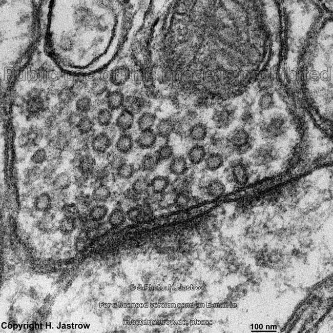
Electron microscopy (EM) offers resolution at the nanometer level, revealing ultrastructural details invisible to light microscopy:
– Synaptic clefts (~20–40 nm wide)
– Synaptic vesicles packed in presynaptic terminals
– Myelin sheaths and nodes of Ranvier
– Mitochondria, ribosomes, and ER
EM is essential for studying synaptic integrity, organelle health, and pathological changes in neurodegenerative diseases.
Drawback: Sample preparation is labor-intensive, and imaging large volumes requires serial sectioning and advanced computational reconstruction.
Recognize Different Neuron Types by Shape
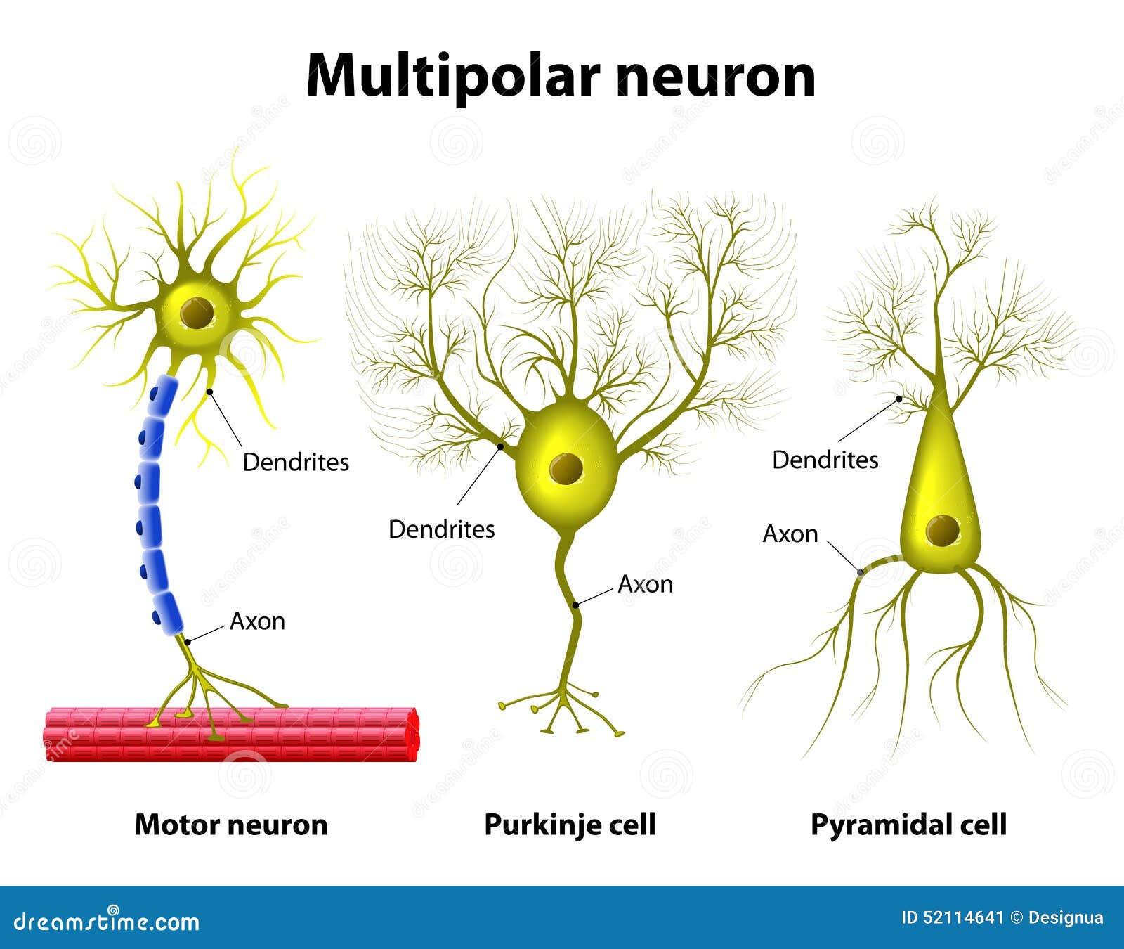
Identify Multipolar Neurons in the Cortex
Multipolar neurons—the most common type—have one axon and multiple dendrites. Examples include:
– Pyramidal cells in the cerebral cortex: triangular soma, a single apical dendrite extending toward the surface, and multiple basal dendrites
– Motor neurons in the spinal cord: large soma with prominent Nissl bodies and long axons
These neurons are typically excitatory, releasing glutamate to activate downstream targets.
Microscopic ID: Look for cells with multiple thick dendrites and a single long axon—common in cortical layers III and V.
Locate Bipolar Neurons in Sensory Pathways
Bipolar neurons have one dendrite and one axon extending from opposite ends of the soma. They are found in:
– Retina: relay visual signals from photoreceptors to ganglion cells
– Olfactory epithelium: transmit smell information to the brain
Under the microscope, they appear as linear cells with processes at both poles.
Function: Enable fast, direct transmission in sensory circuits.
Identify Unipolar (Pseudounipolar) Neurons
Unipolar neurons have a single process that splits into two branches: one acts as a peripheral dendrite (receiving sensory input), and the other as a central axon (sending signals to the spinal cord). Found in dorsal root ganglia (DRG), these neurons transmit touch, pain, and temperature.
Key clue: Look for round cell bodies outside the spinal cord, each with a single fiber that bifurcates.
Note Rare Anaxonic Neurons
Anaxonic neurons lack a clearly defined axon but still transmit signals locally. Found in the retina and olfactory bulb, they are nearly impossible to identify without molecular markers.
Challenge: Their absence of a visible axon makes them indistinguishable in standard histology.
Differentiate Neurons from Glial Cells
Spot Astrocytes in Neural Tissue
Astrocytes are star-shaped glial cells that support neurons by regulating ions, neurotransmitters, and blood flow. Under immunofluorescence with GFAP staining, they appear with elaborate branching processes.
- Small, dark nuclei scattered between neurons
- End feet wrapping around blood vessels
- Critical for forming the blood-brain barrier (BBB)
Warning sign: In injury or disease, astrocytes become reactive, forming glial scars that inhibit regeneration.
Detect Oligodendrocytes and Myelin
Oligodendrocytes are small cells with round nuclei that produce myelin in the CNS. Each can myelinate up to 50 axons.
In Luxol fast blue staining, myelin appears bright blue, revealing white matter tracts.
PNS contrast: Schwann cells myelinate only one axon segment each and are larger and more easily seen.
Observe Microglia as Brain Immune Cells
Microglia are the brain’s resident immune cells. At rest, they have small bodies with long, dynamic processes that constantly survey the tissue.
Upon activation—due to injury, infection, or neurodegeneration—they become amoeboid and migrate to damage sites.
Disease link: Activated microglia are hallmarks of Alzheimer’s, Parkinson’s, and stroke, where they clear debris and release inflammatory signals.
Interpret Synapses and Neural Connectivity
Locate Synapses Under High Magnification
Synapses are visible only under high-resolution imaging:
– Light microscopy: Shows close apposition of axon terminals and dendrites
– Electron microscopy: Reveals:
– Presynaptic terminal with synaptic vesicles
– Synaptic cleft (~30 nm)
– Postsynaptic density with receptor proteins
Excitatory synapses typically occur on dendritic spines, while inhibitory ones target dendritic shafts or soma.
Pro tip: In 3D EM reconstructions, synapses are marked as red dots where axon and dendrite membranes align.
Map Neural Circuits by Cell Type
Neurons are classified by their circuit role:
– Principal neurons (e.g., cortical pyramidal cells): long-range, excitatory
– Interneurons (e.g., chandelier cells): local, mostly inhibitory
Molecular markers like parvalbumin and somatostatin help identify interneuron subtypes.
Functional insight: Inhibitory interneurons control neural rhythms, such as gamma waves essential for attention and memory.
Connect Microscopy to Brain Function and Disease
Microscopic analysis links structure to function:
– Learning → increased spine density
– Memory → spine stabilization
– Neurodegeneration → synapse and neuron loss
Imaging reveals pathology in:
– Alzheimer’s: amyloid plaques, tangles, spine loss
– Multiple sclerosis: demyelinated axons
– Epilepsy: abnormal dendritic sprouting
Future frontier: Connectomics aims to map every neuron and synapse—unlocking the brain’s wiring diagram.
Viewing a neuron under microscope is more than a technical exercise—it’s a window into the biological basis of mind and behavior. From the dark-stained soma to the delicate spines forming trillions of connections, each microscopic detail reflects a functional purpose. Advanced imaging continues to reveal the brain’s dynamic, densely packed reality—where every structure, from axon to astrocyte, plays a role in the symphony of thought.


