
Peering at pollen under a microscope transforms what appears to the naked eye as a fine yellow dust into a breathtaking universe of geometric precision and biological artistry. Pollen under a microscope reveals a hidden world where each grain is a masterfully engineered reproductive package, shaped by millions of years of evolution. These tiny particles—often smaller than a speck of sand—display intricate surface patterns, unique shapes, and specialized structures that reflect the plant’s pollination strategy, whether by wind, insect, bird, or bat.
From the spiky, sticky grains of sunflowers to the smooth, buoyant pollen of pine trees, every feature serves a purpose. Scientists use these microscopic details not only to identify plant species but also to track environmental changes, forecast allergies, solve crimes, and study ancient climates. With magnifications ranging from 100x in basic light microscopes to over 10,000x in electron microscopes, researchers can explore everything from overall shape to nanoscale textures.
This article explores how to prepare and view pollen under a microscope, the key structural features visible at different magnifications, the microscopy techniques that reveal the most detail, and the wide-ranging scientific applications of pollen analysis—from ecology to forensics.
How to Prepare Pollen Slides for Microscopy
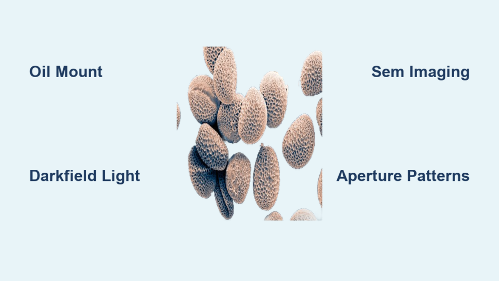
Use Oil Mounts for Clear, Long-Lasting Samples
The best way to view pollen under a microscope is using an oil mount. Place a drop of clear oil—such as sunflower or olive oil—on a clean glass slide. Gently scrape pollen from a flower’s anther using a toothpick and mix it into the oil. Carefully lower a cover slip to avoid trapping air bubbles. Wait a few minutes for the oil to stop flowing before viewing; this ensures stable focus and prevents movement during observation.
Oil prevents pollen grains from absorbing water and bursting, preserving their natural shape. It also enhances transparency, allowing better visualization of internal structures. Slides prepared this way can remain usable for over a year, making them ideal for education or long-term study.
Avoid Water-Based Mounts to Prevent Damage
While water is easily accessible, it’s not ideal for pollen microscopy. Many pollen grains are sensitive to osmotic pressure and will swell, distort, or rupture when placed in water. This destroys delicate surface features like spines and pores, leading to inaccurate observations. If water must be used, examine the sample immediately—within minutes—before degradation occurs.
Dry Mounts Offer Speed but Poor Detail
A dry mount—placing pollen directly on a slide without any medium—is quick but limits image quality. Without a mounting fluid, light doesn’t transmit well through the grains, resulting in dark, low-contrast images. Surface textures are hard to distinguish, and fine details remain hidden. Dry mounts are best reserved for preliminary scans, not detailed analysis.
Enhance Detail with Glycerine and Stains
For high-contrast, professional-grade results, embed pollen in glycerine and add a stain like basic fuchsine. This combination improves cell wall visibility under brightfield or fluorescence microscopy. Stained samples are especially useful for observing internal structures such as the intine and exine layers. While this method requires lab supplies, it delivers superior clarity for research or advanced study.
Microscopy Techniques That Reveal Pollen’s Hidden Details
Brightfield Microscopy Shows Basic Shape and Size
Brightfield microscopy is the most accessible method, using standard transmitted light to reveal pollen shape, size, and general surface patterns at 100x–400x magnification. Grains from Gerbera, Lilium, and dandelion appear clearly defined, making this technique perfect for classrooms or initial observations. Adjusting the light angle with oblique illumination enhances surface texture, bringing out ridges and pores that are otherwise invisible.
Darkfield Microscopy Highlights Surface Textures
Switching to darkfield illumination dramatically increases contrast. By directing light at an angle, the background turns black while the pollen grain glows, making spines, grooves, and apertures stand out. This technique is especially effective for species like poppy (Papaver) and bleeding heart (Lamprocapnos spectabilis), where fine surface features are critical for identification.
Polarized Light Reveals Internal Wall Structure
Polarized light microscopy (PLM) detects birefringent materials in the pollen wall, such as cellulose and sporopollenin. As the slide rotates, structural differences cause light to split, revealing internal layering and symmetry. This method is powerful for identifying pine and sweetgum pollen at 200x–500x and is valuable when surface features are damaged or fossilized.
Scanning Electron Microscopy Captures Nanoscale Topography

Scanning electron microscopy (SEM) produces ultra-high-resolution, 3D-like images of pollen surfaces. Using a focused electron beam, SEM reveals features as small as 10–25 nanometers, including spines, reticulations, viscin threads, and aperture details. Images from USDA ARS of Pinus echinata, Liquidambar styraciflua, and Opuntia stricata showcase this level of detail, with scale bars at 10 µm or 25 µm for accurate size reference. SEM also enables elemental analysis via EDS, useful in forensic and environmental studies.
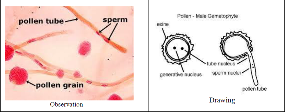
Fluorescence Microscopy Tracks Pollen Tube Growth
Fluorescence microscopy uses dyes like aniline blue to stain callose in growing pollen tubes. Under UV light, the tubes emit a yellowish glow, allowing real-time observation of germination. In Arabidopsis thaliana, this technique visualizes how pollen tubes grow through the style toward the ovule. Inverted images—where tubes appear blue on a white background—are widely used in research publications.
Confocal Microscopy Enables 3D Reconstruction
Confocal laser scanning microscopy captures optical sections at different depths, then stacks them into a 3D model. This is essential for studying pollen tube penetration, intracellular dynamics, or fluorescently labeled proteins. While less common in routine work, confocal imaging is vital in developmental biology and genetic research.
How Pollination Type Shapes Pollen Structure
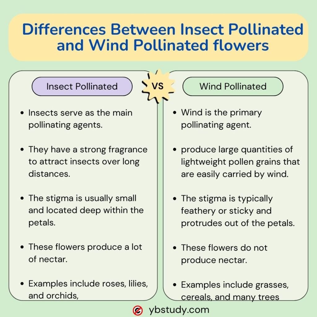
Wind-Pollinated Grains Are Small and Smooth
Plants like pines, oaks, and grasses produce tiny, smooth, lightweight pollen designed for wind dispersal. Pine pollen, for example, has air sacs (bladders) that increase buoyancy, allowing it to travel miles. These grains are typically under 30 µm, spherical or oval, and lack sticky coatings. Their high production volume—billions per tree—compensates for random dispersal and makes them major contributors to seasonal allergies.
Insect-Pollinated Grains Have Spikes and Sticky Coatings
Sunflowers, daisies, and hollyhocks produce larger, textured grains with spines, ridges, and pores to cling to insect bodies. A sticky outer layer called pollenkitt ensures adhesion. Helianthus annuus and Alcea rosea grains are often over 50 µm, with complex sculpting visible under SEM. Some, like Rhododendron, package pollen in tetrads—groups of four—that enhance pollination efficiency.
Bird- and Bat-Pollinated Grains Are Large and Clumped
Bird- and bat-pollinated plants generate large, heavy pollen grains adapted to transfer via feathers or fur. These grains are often sticky or clumped, reducing loss during feeding. Though less studied, they show evolutionary convergence in size and adhesion across distant plant families.
Key Features to Identify in Pollen Grains
Exine Patterns Are Species-Specific
The outer wall—the exine—is made of sporopollenin, one of nature’s most durable materials. Its surface patterns are used to classify species:
– Reticulate: Net-like ridges (Lilium)
– Striate: Parallel lines (Crocus)
– Gemmate: Knobbed texture (Weigela)
– Spiny (echinate): Covered in spikes (Helianthus)
These patterns are visible under SEM or high-magnification light microscopy.
Apertures Indicate Germination Points
Apertures are thin-walled areas where the pollen tube emerges:
– Colpi: Elongated grooves (dicots)
– Pores: Circular openings (monocots)
– Sulci: Single groove (grasses)
Counting and measuring apertures helps classify pollen and infer plant relationships.
Viscin Threads Bind Pollen into Functional Units
Found in Ericaceae and Onagraceae, viscin threads are sticky filaments that bind pollen into bundles. This ensures multiple grains are transferred at once, increasing fertilization success. Visible under high magnification, they appear as fine, web-like strands.
Tetrads Reveal Meiotic Packaging
In some species, pollen remains in groups of four after meiosis. Common in Rhododendron, tetrads provide insight into microsporogenesis and reproductive biology.
Applications of Pollen Microscopy in Science
Track Biodiversity with Automated Imaging
The Pollen Microscope at Massey University captures nine focal planes per grain, creating 3D reconstructions to measure volume, surface area, and texture. It processes hundreds of grains per hour, enabling rapid classification by AI and supporting ecological monitoring.
Map Pollination Networks via Bee Pollen
CSIRO analyzes pollen from bee corbiculae using automated microscopy to map pollination networks, assess ecosystem health, and support sustainable agriculture.
Reconstruct Ancient Climates from Fossil Pollen
Palynologists extract fossil pollen from sediment cores to reconstruct past vegetation and climate. Shifts in pollen assemblages reveal changes in temperature, rainfall, and land use.
Forecast Allergies Using Airborne Counts
Public health agencies measure daily pollen counts to predict hay fever outbreaks. In Atlanta, levels over 6,974 grains/m³ correlate with widespread symptoms. Top allergens include pine, oak, mulberry, sweetgum, and birch.
Solve Crimes with Forensic Palynology
Pollen’s durability and species specificity make it a powerful geolocational biomarker. Grains on clothing or vehicles can link suspects to crime scenes.
Verify Honey Origin in Apiculture
Beekeepers analyze pollen in honey to verify floral sources, ensuring authenticity in products labeled as “wildflower” or “orange blossom.”
Final Note: Pollen under a microscope is more than a scientific curiosity—it’s a gateway to understanding life’s interconnectedness. From allergy forecasts to ancient climate records, each grain carries a story written in shape, texture, and chemistry. Whether you’re a student, researcher, or nature enthusiast, observing pollen reveals the astonishing precision of nature’s smallest messengers.


