
A single drop of pond water holds a hidden universe. Zoom in with a microscope, and you’ll witness protozoa under microscope—tiny, living dramas unfolding in real time. These single-celled eukaryotes dart, glide, and pulse with purpose, each movement revealing a sophisticated survival strategy. From the slow ooze of an amoeba to the lightning strike of a predatory Didinium, protozoa offer a front-row seat to the complexity of life at the microscopic scale.
Observing protozoa under microscope is more than a lab exercise—it’s a gateway to understanding ecology, cellular biology, and the unseen forces shaping soil fertility and water health. With just a compound microscope (100x–400x magnification), a wet mount slide, and a sample from a nearby pond or compost, you can explore this dynamic world. This guide walks you through the major groups, how to identify them, what behaviors to watch for, and why they matter—so you can turn curiosity into discovery.
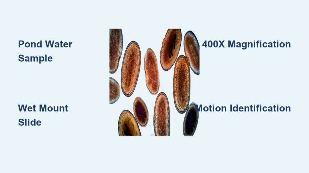
Spotting Protozoa: Where and How to Look
Finding protozoa begins with knowing the right environments and techniques. These microbes thrive in moist habitats rich in bacteria—their primary food source. A drop of water from a stagnant pond, ditch, or even soaked garden soil can teem with dozens of species if observed quickly.
Best Sample Sources
- Pond or ditch water – especially near decaying leaves or sediment, where microbial activity is high.
- Compost tea or soil extract – mix garden soil with distilled water, let it settle, then pipette the upper layer for motile organisms.
- Hay infusions – soak dried grass in water for 2–3 days; this encourages bacterial blooms and attracts protozoa.
Microscope Setup Tips
- Use a compound light microscope with 40x, 100x, and 400x objectives.
- Prepare a wet mount slide: place a drop of sample on a clean slide, gently lower a cover slip to avoid bubbles.
- Slightly close the iris diaphragm to reduce light and increase contrast—critical for transparent organisms.
- Observe within minutes—protozoa lose motility when dried or exposed to air.
What Movement Tells You
- Slow, flowing motion → likely an amoeba.
- Fast, jerky, or spiral movement → probably a flagellate.
- Smooth, rotating glide → almost certainly a ciliate.
- Sudden contraction → look for Vorticella or Carchesium on stalks.
These movement patterns are your first clues to identification, even before you see detailed structures.
Amoebae: Masters of Shape-Shifting Motion
Amoebae move by extending pseudopodia—”false feet” formed by flowing cytoplasm. This amoeboid motion allows them to crawl across surfaces and engulf prey with surprising precision.
How to Identify Amoebae
- Irregular, changing shape – no fixed form.
- Transparent, jelly-like body – best seen with reduced light.
- Slow gliding across the slide.
- Visible food vacuoles after eating bacteria or other microbes.
- Nucleus visible in larger species like Amoeba proteus.
Watch for Phagocytosis in Action
When an amoeba encounters prey like Paramecium or bacteria, it engulfs it via phagocytosis. You’ll see:
– Cytoplasm flow around the target.
– Formation of a food vacuole inside the cell.
– Vacuole circulate as digestion occurs.
Amoeba proteus can grow up to 500 micrometers—visible even at 100x—and has been observed consuming Paramecium containing green algae, showing multi-level feeding.
Testate Amoebae: The Shelled Protozoa
Some amoebae build protective shells:
– Difflugia: flask-shaped test made of sand grains.
– Arcella: dome-like, helmet-shaped shell.
Both extend pseudopodia through an opening to move and feed. Found in pond sediments and peat bogs.
Flagellates: The Speedsters of the Micro World
Flagellates use one or more whip-like flagella to propel themselves through water. Their movement is fast, darting, and often spiral—making them stand out in a crowded field.
Key Features to Spot
- Elongated or oval cell shape.
- Rapid, directional motion—they respond quickly to light or chemicals.
- Single flagellum (e.g., Euglena) or multiple flagella.
- At 400x, flagella may be visible as thin threads.
Photosynthetic Flagellates
Some flagellates perform photosynthesis:
– Euglena: green due to chloroplasts, with a red eyespot to detect light.
– Ugina gracilis: contains chloroplasts and a pigment shield for phototaxis.
These often cluster near light sources in a culture.
Identification Clues
- Look for jerky forward bursts followed by brief pauses.
- Best seen in fresh, undisturbed samples—they die quickly when dried.
- Often appear smaller than ciliates (15–50 µm).
Ciliates: The Complex Giants of Protozoa
Ciliates are the largest and most complex protozoa, covered in hundreds to thousands of cilia that beat in coordinated waves. They move with smooth, rotating precision and often dominate pond samples.
How to Recognize Ciliates
- Large size (50–300 µm, some over 1 mm).
- Dense, shimmering coat of cilia—looks like a vibrating surface.
- Spiral or helical swimming path.
- Oral groove visible in some species (e.g., Paramecium).
Watch for Feeding Activity
- Cytostome (gullet) draws in bacteria via water currents.
- Food vacuoles form and circulate—can be tracked with dyes like carmine red.
- Undigested waste expelled through cytoproct (anal pore).
Common Ciliates & Their Traits
Paramecium: The Slipper-Shaped Hunter
- Elongated, slipper-like shape.
- Moves via ~10,000 cilia in metachronal waves.
- Forms food vacuoles; expels waste.
- Undergoes conjugation—two individuals exchange genetic material.
Didinium: The Ciliate Assassin
- Predatory ciliate that hunts Paramecium.
- Fires protein-coiled harpoons (pistils) to immobilize prey.
- Swallows entire Paramecium, which remains visible inside.
Vorticella: The Bell on a Spring
- Bell-shaped cell attached by a contractile stalk.
- Stalk contains myoneme fibers—contracts in milliseconds when disturbed.
- Looks like a tiny flower that snaps shut.
Carchesium: Colonial Vorticella
- Multiple Vorticella-like cells on branching stalks.
- Entire colony expands and contracts together—a rare example of synchronized behavior in unicells.
Tetrahymena: Model Organism
- Smaller than Paramecium (40–70 µm).
- Used in genetics and cell biology research.
- Reproduces via binary fission and conjugation.
Other Fascinating Protozoa to Watch For
Beyond the big three groups, several other protozoa reveal unique adaptations.
Heliozoa: The Sun Animalcules
- Spherical with radiating axopodia (rigid, sticky projections).
- Traps bacteria and flagellates on its “spikes.”
- Upon contact, axopodia rapidly collapse to pull prey in.
- Actinophrys: common in pond water; best seen with DIC microscopy.
Dinoflagellates: Marine Killers
- Mostly marine; two flagella (one equatorial, one longitudinal).
- Some cause red tides and produce deadly toxins.
- Under microscope: armored cells with grooves; some bioluminescent.
Colonial & Giant Protozoa
Volvox: The Rolling Green Sphere
- Colonial alga with hundreds of flagellated cells.
- Each cell has two flagella and an eyespot.
- Daughter colonies form inside the parent sphere.
- Visible at 100x; rolls smoothly through water.
Spirostomum: The Giant Unicell
- One of the largest known single cells—over 1 mm long.
- Elongated, worm-like, but entirely unicellular.
- Contracts rapidly when disturbed.
- Glides using cilia; resembles a nematode but isn’t.
Microscopy Techniques for Clear Viewing
Even basic equipment can reveal stunning detail—if used correctly.
Essential Tools
- Compound microscope (40x–400x).
- Glass slides and cover slips.
- Pipette for sample transfer.
- Iris diaphragm to adjust contrast.
Wet Mount Preparation
- Place a drop of sample on a clean slide.
- Gently lower a cover slip at an angle to avoid bubbles.
- If too dry, add a tiny drop at the edge.
- Observe immediately—motility fades fast.
Boosting Visibility
- Close the iris diaphragm 50–75% to increase contrast.
- Use phase contrast or DIC if available—reveals cilia, flagella, nuclei.
- Staining (e.g., methylene blue) highlights nuclei but kills live cells.
Magnification Guide
| Magnification | Best For |
|---|---|
| 40x | Scanning; spotting large ciliates, rotifers |
| 100x | General ID: Paramecium, Amoeba, Volvox |
| 400x | Detail: cilia, flagella, food vacuoles, phagocytosis |
Why Protozoa Matter: Ecology & Soil Health
Protozoa aren’t just fascinating—they’re ecosystem engineers.
Nutrient Cycling Powerhouses
- Feed on bacteria, releasing ammonium (NH₄⁺).
- Up to 80% of nitrogen mineralization in soil is protozoan-driven.
- Enhances plant nutrient availability naturally.
Soil Health Indicators
| Protozoan Group | Soil Condition |
|---|---|
| Amoebae | Aerated, rich in organic matter |
| Flagellates | Moist, biologically active |
| Ciliates | Waterlogged, compacted, or anaerobic |
High protozoan diversity = healthy, fertile soil.
Absence may signal over-tillage, chemical toxicity, or poor aeration.
Microbial Balance
- Prevent bacterial overgrowth.
- Promote microbial diversity and resilience.
- Influence fungal:bacterial ratios in soil food webs.
Quick ID Chart: Protozoa You’ll Likely See
| Organism | Locomotion | Size | Key Feature |
|---|---|---|---|
| Amoeba proteus | Pseudopodia | Up to 500 µm | Shape-shifting, phagocytosis |
| Paramecium | Cilia | 50–300 µm | Slipper-shaped, oral groove |
| Euglena | Flagellum | 15–50 µm | Green, red eyespot |
| Vorticella | Cilia (stalked) | 30–100 µm | Bell-shaped, contracts fast |
| Didinium | Cilia | 50–150 µm | Predatory, harpoons Paramecium |
| Volvox | Flagella (colonial) | 300–500 µm | Rolling green sphere |
| Difflugia | Pseudopodia | 100–200 µm | Sand-grain shell |
| Spirostomum | Cilia | >1 mm | Giant, worm-like unicell |
With just a microscope and a drop of water, you can explore a world teeming with life. Protozoa under microscope reveal not only biological complexity but also ecological connections that sustain our planet. Whether you’re a student, gardener, or curious observer, these “micro friends” invite you to see the unseen—and appreciate the drama of life at the smallest scale.


