
Have you ever wondered how your body moves food through the digestive tract, regulates blood flow, or controls bladder emptying—without you even thinking about it? The answer lies in smooth muscle, an involuntary tissue that works behind the scenes to maintain essential bodily functions. When viewed under a microscope, smooth muscle stands out due to its spindle-shaped cells, single central nucleus, and lack of striations—features that make it easily distinguishable from skeletal and cardiac muscle. Whether you’re analyzing a blood vessel, intestinal wall, or uterine tissue, recognizing smooth muscle is crucial for understanding both normal physiology and disease processes. In this guide, you’ll learn how to confidently identify smooth muscle under the microscope, interpret its appearance in different orientations, and differentiate it from similar tissues using histological clues and staining techniques.
Identify Key Microscopic Features
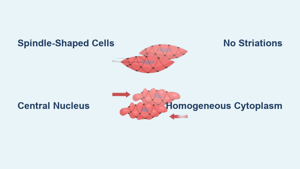
Recognize Spindle-Shaped Cell Morphology
One of the most defining traits of smooth muscle is its fusiform (spindle-shaped) cells, which are thick in the center and taper at both ends—resembling a cigar or kayak. In longitudinal sections, these elongated cells often appear wavy or staggered due to natural resting tension and overlapping arrangement. Their length ranges from 10 to 200 microns, depending on location and function. Because the cells are tightly packed and connected by gap junctions, their borders are frequently indistinct, creating a woven or bundled appearance. This unique shape allows for multidirectional contraction, essential for organs like the intestines and uterus.
Locate the Central Nucleus
Each smooth muscle cell contains one elongated nucleus positioned centrally. In longitudinal view, the nucleus aligns with the long axis of the cell and appears cigar-shaped or ovoid. A helpful diagnostic clue is that during contraction, the nucleus may kink, twist, or spiral, reflecting the cell’s shortening. In cross-sections, only cells cut through the midsection show a visible nucleus—appearing as a central dot or oval—while those sectioned peripherally lack nuclei entirely. This mix of nucleated and anucleate profiles is a key feature when identifying smooth muscle in cross-sectional views.
Confirm Absence of Striations
Unlike skeletal and cardiac muscle, smooth muscle lacks sarcomeres, the repeating contractile units responsible for striations. As a result, its cytoplasm appears homogeneous and smooth under light microscopy. There are no alternating dark (A) and light (I) bands, Z-lines, or M-lines. Even at high magnification with routine H&E staining, the cytoplasm remains uniformly pink without any banding pattern. This absence of striations is so characteristic that it gives the tissue its name—smooth muscle.
Analyze Sectioning Effects on Appearance
![]()
Interpret Longitudinal Sections Correctly
In longitudinal sections, smooth muscle fibers run parallel to the plane of sectioning. You’ll typically see:
– Elongated, overlapping cells with indistinct membranes
– Central, cigar-shaped nuclei aligned along the fiber axis
– Homogeneous, acidophilic (pink) cytoplasm
– A wavy or zig-zag pattern caused by natural contraction or tissue fixation
The staggered arrangement supports synchronized contraction across large sheets, especially in organs like the intestine and bladder. However, due to overlapping, individual cell boundaries can be hard to trace—requiring careful observation of nuclear alignment and shape.
Decode Cross-Sectional Views
Cross-sections reveal smooth muscle as a tightly packed field of circular or polygonal profiles, resembling a honeycomb. The appearance depends on where the plane intersects the fusiform cell:
– Central cuts show a nucleus in the middle (dot or oval)
– Peripheral cuts appear as anuclear, eosinophilic rings with no visible nucleus
This variation in nuclear visibility helps distinguish smooth muscle from dense connective tissue, which typically lacks organized cellular profiles and shows sparse, flattened fibroblasts.
Compare Smooth vs. Other Muscle Types
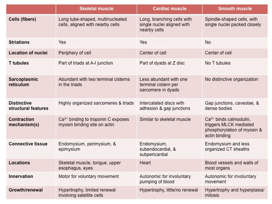
Differentiate from Skeletal Muscle
Skeletal muscle is strikingly different from smooth muscle under the microscope:
– Striations: Present in skeletal, absent in smooth
– Cell shape: Cylindrical and long (skeletal) vs. spindle-shaped (smooth)
– Nuclei: Multiple, peripherally located (skeletal) vs. single, central (smooth)
– Control: Voluntary (skeletal) vs. involuntary (smooth)
In tissues like the esophagus, both types coexist—outer layer skeletal, inner layer smooth—providing a direct comparison in one histological slide.
Distinguish from Cardiac Muscle
While cardiac muscle shares some features with smooth muscle (e.g., involuntary control, central nuclei), key differences eliminate confusion:
– Striations: Present in cardiac, absent in smooth
– Cell branching: Unique to cardiac tissue
– Intercalated discs: Dark, stair-step junctions visible with staining
– Nuclei: One or two central nuclei per cell, not elongated
These features are especially important when examining vascular or heart tissue sections.
Use Staining Techniques for Confirmation
Apply H&E Staining Basics
The standard hematoxylin and eosin (H&E) stain is the first step in identifying smooth muscle:
– Cytoplasm: Stains pink (acidophilic) due to high actin and myosin content
– Nuclei: Stain blue-purple with hematoxylin
– Background: Uniformly pink without banding
However, since collagen also stains pink, H&E alone can lead to misidentification in dense tissues. Always look for cellular organization and nuclear position to confirm.
Employ Special Stains for Clarity
When H&E is inconclusive, special stains provide definitive differentiation:
– Mallory trichrome: Smooth muscle → Red, collagen → Blue
– Masson’s trichrome: Muscle → red, collagen → green/blue
– Verhoeff-Van Gieson (elastin stain): Highlights elastic fibers in vessel walls
– Silver stains (Bodian method): Reveal the basal lamina as a dotted or interrupted line around each cell, confirming individual boundaries
These stains are invaluable in diagnosing tumors (e.g., distinguishing leiomyoma from fibroma) or analyzing vascular pathology.
Understand Functional Tissue Arrangement

Observe Layered Organization in Organs
Smooth muscle is organized into functional layers that enable coordinated movement:
– Gastrointestinal tract:
– Inner circular layer → constricts lumen
– Outer longitudinal layer → shortens organ
– Together, they generate peristaltic waves
– Uterus (myometrium): Three interwoven layers allow multidirectional contraction during labor
– Blood vessels: Circular arrangement regulates diameter via vasoconstriction/vasodilation
This layered architecture is clearly visible in cross-sectional slides of arteries and intestines.
Recognize Sphincter and Circular Band Structures
Specialized smooth muscle forms ring-like sphincters that control passage:
– Lower esophageal sphincter
– Pyloric sphincter
– Internal anal and urethral sphincters
These appear as thickened circular bundles in histology, often surrounded by connective tissue. Their involuntary nature contrasts with voluntary external sphincters composed of skeletal muscle.
Locate Common Body Sites
Find Smooth Muscle in GI Tract
The digestive system is rich in smooth muscle:
– Stomach, small intestine, colon: Double-layered muscularis externa (circular + longitudinal)
– Muscularis mucosae: Thin inner layer beneath epithelium—visible as a fine pink band in colon biopsies
– Internal anal sphincter: Smooth muscle continuation of circular layer
In histology slides, look for pink, layered tissue beyond the submucosa.
Identify Vascular and Respiratory Locations
- Arteries and arterioles: Thick smooth muscle walls regulate blood pressure
- Muscular arteries show swirling layers in cross-section
- Veins and lymphatics: Thinner muscle layer but still identifiable
- Bronchi and bronchioles: Circular smooth muscle controls airflow
- Contraction → bronchoconstriction (e.g., in asthma)
In vessel walls, smooth muscle blends with elastic fibers—use elastin stains to separate them.
Spot Urogenital and Specialized Sites
- Uterus (myometrium): Thick, interlacing bundles; hypertrophy during pregnancy
- Bladder (detrusor muscle): Three loosely organized layers; contracts to expel urine
- Ureters, vas deferens: Propel fluids via peristalsis
- Iris (sphincter pupillae) and ciliary body: Control pupil size and lens shape
- Arrector pili muscles: Attach to hair follicles; cause “goosebumps”
These locations highlight smooth muscle’s role in both homeostasis and reflex responses.
Avoid Common Identification Mistakes
Don’t Confuse with Dense Connective Tissue
Both smooth muscle and collagen stain pink with H&E, leading to frequent misidentification. Key differences:
– Smooth muscle: Uniform spindle cells with central nuclei in longitudinal view
– Dense connective tissue: No cells in cross-section; fibroblasts are sparse and flattened
Use trichrome stains (Mallory or Masson’s) to resolve ambiguity: red = muscle, blue/green = collagen.
Watch for Sectioning Artifacts
Oblique or tangential cuts can distort appearance:
– Longitudinal sections may show partial nuclei or irregular shapes
– Cross-sections may mimic fat or cartilage if not properly oriented
Always examine multiple fields and use consistent identifiers: fusiform shape, central nucleus, no striations.
Master 3D Interpretation from 2D Slides
Reconstruct Cellular Shape Across Sections
Histological sections are 2D slices of 3D tissue. A single cell may appear:
– Long and spindle-shaped (longitudinal)
– Round with a nucleus (central cross-section)
– Small and anuclear (peripheral cut)
Recognizing this variability prevents misinterpretation. True identification comes from pattern recognition across multiple planes.
Use Nuclear Position as a Guide
The central nucleus is a reliable landmark:
– If nuclei are centrally aligned in elongated cells → likely smooth muscle
– If nuclei are peripheral → likely skeletal muscle
– If nuclei are central but striations present → cardiac muscle
This visual cue speeds up accurate identification.
Summary: Quick Identification Checklist
Core Features to Confirm
When viewing a slide, ask:
– Are the cells spindle-shaped?
– Is there a single, central nucleus?
– Are no striations visible?
– Is the cytoplasm homogeneously pink?
– Are cells arranged in sheets or bundles?
If yes to all, it’s smooth muscle.
Staining and Context Tips
- Use trichrome stains to rule out collagen
- Check location: GI tract, blood vessels, uterus?
- Compare with adjacent tissues for contrast
- Look for functional structures like sphincters or vessel walls
Smooth muscle is a cornerstone of involuntary bodily functions, and its histological identification is fundamental in anatomy, pathology, and clinical diagnostics. By mastering its appearance in both longitudinal and cross-sections, applying special stains when needed, and understanding its functional organization, you can confidently distinguish smooth muscle from other tissues. Whether you’re studying peristalsis, vascular tone, or reproductive physiology, recognizing this tissue type enhances your ability to interpret biological processes at the microscopic level.


